 Movie
Movie Controller

[English] 日本語
 Yorodumi
- PDB-1kuy: X-ray Crystallographic Studies of Serotonin N-acetyltransferase C... -
+ Open data

- Basic information
| Entry | Database: PDB / ID: 1kuy | ||||||
|---|---|---|---|---|---|---|---|
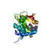
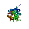
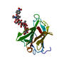
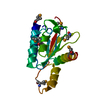
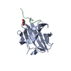
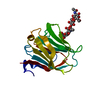
| Title | X-ray Crystallographic Studies of Serotonin N-acetyltransferase Catalysis and Inhibition | ||||||
 Components Components | Serotonin N-acetyltransferase Aralkylamine N-acetyltransferase Aralkylamine N-acetyltransferase | ||||||
 Keywords Keywords | TRANSFERASE / Enzyme-Inhibitor Complex / Bisubstrate Analog / Alternate Conformations | ||||||
| Function / homology |  Function and homology informationaralkylamine N-acetyltransferase / melatonin biosynthetic process / aralkylamine N-acetyltransferase activity / N-terminal protein amino acid acetylation / cellular response to cAMP / circadian rhythm / perinuclear region of cytoplasm Function and homology informationaralkylamine N-acetyltransferase / melatonin biosynthetic process / aralkylamine N-acetyltransferase activity / N-terminal protein amino acid acetylation / cellular response to cAMP / circadian rhythm / perinuclear region of cytoplasmSimilarity search - Function | ||||||
| Biological species |  Ovis aries (sheep) Ovis aries (sheep) | ||||||
| Method | X-RAY DIFFRACTION / MOLECULAR REPLACEMENT / Resolution: 2.4 Å | ||||||
 Authors Authors | Wolf, E. / De Angelis, J. / Khalil, E.M. / Cole, P.A. / Burley, S.K. | ||||||
 Citation Citation | Journal: J.Mol.Biol. / Year: 2002 Title: X-ray crystallographic studies of serotonin N-acetyltransferase catalysis and inhibition. Authors: Wolf, E. / De Angelis, J. / Khalil, E.M. / Cole, P.A. / Burley, S.K. | ||||||
| History |
|
- Structure visualization
| Structure viewer | Molecule: MolmilJmol/JSmol |
|---|
- Downloads & links
-Download
| PDBx/mmCIF format | 1kuy.cif.gz | 51.6 KB | Display | PDBx/mmCIF format |
|---|---|---|---|---|
| PDB format | pdb1kuy.ent.gz | 36.2 KB | Display | PDB format |
| PDBx/mmJSON format | 1kuy.json.gz | Tree view | PDBx/mmJSON format | |
| Others |  Other downloads Other downloads |
-Validation report
| Arichive directory | https://data.pdbj.org/pub/pdb/validation_reports/ku/1kuyftp://data.pdbj.org/pub/pdb/validation_reports/ku/1kuy | HTTPS FTP |
|---|
-Related structure data
| Related structure data | 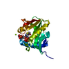 1kuvSC 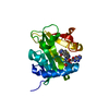 1kuxC S: Starting model for refinement C: citing same article ( |
|---|---|
| Similar structure data |


-Links
 PDBj
PDBj

- Assembly
| Deposited unit | 
| ||||||||||
|---|---|---|---|---|---|---|---|---|---|---|---|
| 1 |
| ||||||||||
| Unit cell |
| ||||||||||
| Details | The biological assembly is a monomer |
-Components
| #1: Protein | Aralkylamine N-acetyltransferase / E.C.2.3.1.87 / arylalkylamine N-acetyltransferase / Aralkylamine N-acetyltransferase / AA-NAT / Serotonin acetylase Mass: 23111.568 Da / Num. of mol.: 1 / Mutation: MET substituted by Se-met Source method: isolated from a genetically manipulated source Source: (gene. exp.) Ovis aries (sheep) / Gene: U29663 / Plasmid: pGEX-6P-1 / Production host:  Escherichia coli (E. coli) / Strain (production host): BL21(DE3)pLysS Escherichia coli (E. coli) / Strain (production host): BL21(DE3)pLysSReferences: UniProt: Q29495, aralkylamine N-acetyltransferase |
|---|---|
| #2: Chemical | ChemComp-COT /   Mass: 967.771 Da / Num. of mol.: 1 / Source method: obtained synthetically / Formula: C33H48N9O17P3S Mass: 967.771 Da / Num. of mol.: 1 / Source method: obtained synthetically / Formula: C33H48N9O17P3S |
| #3: Water | ChemComp-HOH / Water Mass: 18.015 Da / Num. of mol.: 74 / Source method: isolated from a natural source / Formula: H2O Mass: 18.015 Da / Num. of mol.: 74 / Source method: isolated from a natural source / Formula: H2O |
-Experimental details
-Experiment
| Experiment | Method: X-RAY DIFFRACTION / Number of used crystals: 1 |
|---|
- Sample preparation
| Crystal grow | Temperature: 277 K / Method: vapor diffusion, hanging drop / pH: 6.5 Details: PEG 2000, MPD, ammonium sulfate, MES pH 6.5, magnesium acetate, DTT, spermidine, and lithium chloride. VAPOR DIFFUSION, HANGING DROP at 277K, temperature 277.0K | ||||||||||||||||||||||||||||||||||||||||||||||||||||||||||||||||||||||
|---|---|---|---|---|---|---|---|---|---|---|---|---|---|---|---|---|---|---|---|---|---|---|---|---|---|---|---|---|---|---|---|---|---|---|---|---|---|---|---|---|---|---|---|---|---|---|---|---|---|---|---|---|---|---|---|---|---|---|---|---|---|---|---|---|---|---|---|---|---|---|---|
| Crystal grow | *PLUS Temperature: 4 ℃ / Details: used microseeding | ||||||||||||||||||||||||||||||||||||||||||||||||||||||||||||||||||||||
| Components of the solutions | *PLUS
|
-Data collection
| Diffraction | Mean temperature: 100 K |
|---|---|
| Diffraction source | Source: ROTATING ANODE / Type: RIGAKU RU200 / Wavelength: 1.5418 Å |
| Detector | Type: RIGAKU RAXIS IIC / Detector: IMAGE PLATE / Date: Apr 9, 2000 / Details: Yale mirrors |
| Radiation | Monochromator: Coated Yale mirrors, 0.00015" Nickel filter / Protocol: SINGLE WAVELENGTH / Monochromatic (M) / Laue (L): M / Scattering type: x-ray |
| Radiation wavelength | Wavelength: 1.5418 Å / Relative weight: 1 |
| Reflection | Resolution: 2.4→20 Å / Num. all: 6689 / Num. obs: 6248 / % possible obs: 93.4 % / Observed criterion σ(I): -3 / Redundancy: 3.7 % / Rmerge(I) obs: 0.088 / Rsym value: 0.088 / Net I/σ(I): 17.4 |
| Reflection shell | Resolution: 2.4→2.49 Å / Redundancy: 3 % / Rmerge(I) obs: 0.133 / Mean I/σ(I) obs: 12.3 / Num. unique all: 650 / Rsym value: 0.133 / % possible all: 94.2 |
| Reflection | *PLUS Highest resolution: 2.4 Å / Lowest resolution: 20 Å |
| Reflection shell | *PLUS % possible obs: 94.2 % |
- Processing
| Software |
| |||||||||||||||||||||||||
|---|---|---|---|---|---|---|---|---|---|---|---|---|---|---|---|---|---|---|---|---|---|---|---|---|---|---|
| Refinement | Method to determine structure: MOLECULAR REPLACEMENT Starting model: 1KUV Resolution: 2.4→20 Å / Cross valid method: THROUGHOUT / σ(F): 0 / σ(I): 0 / Stereochemistry target values: Engh & Huber Details: Structure was refined with waters and ligand was modeled based on difference fourier electron density. Ligand was included in final refinement
| |||||||||||||||||||||||||
| Displacement parameters |
| |||||||||||||||||||||||||
| Refinement step | Cycle: LAST / Resolution: 2.4→20 Å
| |||||||||||||||||||||||||
| Refine LS restraints |
| |||||||||||||||||||||||||
| Refinement | *PLUS Highest resolution: 2.4 Å / Lowest resolution: 20 Å / % reflection Rfree: 10 % / Rfactor obs: 0.203 | |||||||||||||||||||||||||
| Solvent computation | *PLUS | |||||||||||||||||||||||||
| Displacement parameters | *PLUS | |||||||||||||||||||||||||
| Refine LS restraints | *PLUS Type: c_angle_deg / Dev ideal: 1.2 |
