 Movie
Movie Controller

[English] 日本語
 Yorodumi
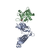
- PDB-1ktk: Complex of Streptococcal pyrogenic enterotoxin C (SpeC) with a hu... -
+ Open data

- Basic information
| Entry | Database: PDB / ID: 1ktk | ||||||
|---|---|---|---|---|---|---|---|
| Title | Complex of Streptococcal pyrogenic enterotoxin C (SpeC) with a human T cell receptor beta chain (Vbeta2.1) | ||||||
 Components Components |
| ||||||
 Keywords Keywords |  IMMUNE SYSTEM / Streptococcus / immunity / T cell receptor beta IMMUNE SYSTEM / Streptococcus / immunity / T cell receptor beta | ||||||
| Function / homology |  Function and homology information Function and homology informationalpha-beta T cell receptor complex / Translocation of ZAP-70 to Immunological synapse / Phosphorylation of CD3 and TCR zeta chains / Generation of second messenger molecules / alpha-beta T cell activation / PD-1 signaling / Immunoregulatory interactions between a Lymphoid and a non-Lymphoid cell / Downstream TCR signaling / T cell receptor signaling pathway / toxin activity ...alpha-beta T cell receptor complex / Translocation of ZAP-70 to Immunological synapse / Phosphorylation of CD3 and TCR zeta chains / Generation of second messenger molecules / alpha-beta T cell activation / PD-1 signaling / Immunoregulatory interactions between a Lymphoid and a non-Lymphoid cell / Downstream TCR signaling / T cell receptor signaling pathway / toxin activity / adaptive immune response / immune response / extracellular region / membrane / plasma membraneSimilarity search - Function | ||||||
| Biological species |  Streptococcus pyogenes (bacteria) Streptococcus pyogenes (bacteria) Homo sapiens (human) Homo sapiens (human) | ||||||
| Method | X-RAY DIFFRACTION / SYNCHROTRON / Resolution: 3 Å | ||||||
 Authors Authors | Sundberg, E.J. / Li, H. / Llera, A.S. / McCormick, J.K. / Tormo, J. / Karjalainen, K. / Schlievert, P.M. / Mariuzza, R.A. | ||||||
 Citation Citation | Journal: Structure / Year: 2002 Title: Structures of two streptococcal superantigens bound to TCR beta chains reveal diversity in the architecture of T cell signaling complexes. Authors: Sundberg, E.J. / Li, H. / Llera, A.S. / McCormick, J.K. / Tormo, J. / Schlievert, P.M. / Karjalainen, K. / Mariuzza, R.A. | ||||||
| History |
|
- Structure visualization
| Structure viewer | Molecule: MolmilJmol/JSmol |
|---|
- Downloads & links
-Download
| PDBx/mmCIF format | 1ktk.cif.gz | 241.7 KB | Display | PDBx/mmCIF format |
|---|---|---|---|---|
| PDB format | pdb1ktk.ent.gz | 203 KB | Display | PDB format |
| PDBx/mmJSON format | 1ktk.json.gz | Tree view | PDBx/mmJSON format | |
| Others |  Other downloads Other downloads |
-Validation report
| Arichive directory | https://data.pdbj.org/pub/pdb/validation_reports/kt/1ktkftp://data.pdbj.org/pub/pdb/validation_reports/kt/1ktk | HTTPS FTP |
|---|
-Related structure data


-Links
 PDBj
PDBj


- Assembly
| Deposited unit | 
| ||||||||||
|---|---|---|---|---|---|---|---|---|---|---|---|
| 1 |
| ||||||||||
| Unit cell |
|
-Components
| #1: Protein | Mass: 24384.285 Da / Num. of mol.: 4 / Mutation: H35A Source method: isolated from a genetically manipulated source Source: (gene. exp.) Streptococcus pyogenes (bacteria) / Production host: Escherichia coli (E. coli) / References: UniProt: P13380, UniProt: Q8NKX2*PLUS#2: Protein | Mass: 27461.572 Da / Num. of mol.: 2 / Mutation: C13A, C191A Source method: isolated from a genetically manipulated source Source: (gene. exp.) Homo sapiens (human) / Production host: Escherichia coli (E. coli) / References: UniProt: P01850 |
|---|
-Experimental details
-Experiment
| Experiment | Method: X-RAY DIFFRACTION / Number of used crystals: 1 |
|---|
- Sample preparation
| Crystal | Density Matthews: 3.7 Å3/Da / Density % sol: 66.79 % | |||||||||||||||||||||||||||||||||||
|---|---|---|---|---|---|---|---|---|---|---|---|---|---|---|---|---|---|---|---|---|---|---|---|---|---|---|---|---|---|---|---|---|---|---|---|---|
| Crystal grow | Temperature: 298 K / Method: vapor diffusion, hanging drop / pH: 8 Details: 15% PEG 8000, 0.2 M MgCl2, pH 8.0, VAPOR DIFFUSION, HANGING DROP at 298K | |||||||||||||||||||||||||||||||||||
| Crystal grow | *PLUS | |||||||||||||||||||||||||||||||||||
| Components of the solutions | *PLUS
|
-Data collection
| Diffraction | Mean temperature: 100 K |
|---|---|
| Diffraction source | Source: SYNCHROTRON / Site: CHESS  / Beamline: A1 / Wavelength: 0.978 Å / Beamline: A1 / Wavelength: 0.978 Å |
| Detector | Type: ADSC QUANTUM 4 / Detector: CCD / Date: Sep 8, 2000 |
| Radiation | Protocol: SINGLE WAVELENGTH / Monochromatic (M) / Laue (L): M / Scattering type: x-ray |
| Radiation wavelength | Wavelength: 0.978 Å / Relative weight: 1 |
| Reflection | Resolution: 3→100 Å / Num. all: 43729 / Num. obs: 36549 / % possible obs: 94.1 % / Observed criterion σ(F): 0 / Observed criterion σ(I): 0 |
| Reflection shell | Resolution: 3→3.16 Å / % possible all: 81.9 |
| Reflection | *PLUS Highest resolution: 3 Å / Lowest resolution: 100 Å / Num. obs: 43729 / % possible obs: 97.8 % / Num. measured all: 158446 / Rmerge(I) obs: 0.05 |
| Reflection shell | *PLUS % possible obs: 96.5 % / Rmerge(I) obs: 0.207 / Mean I/σ(I) obs: 6.5 |
- Processing
| Software |
| ||||||||||||||||||||
|---|---|---|---|---|---|---|---|---|---|---|---|---|---|---|---|---|---|---|---|---|---|
| Refinement | Resolution: 3→6 Å / Rfactor Rfree error: 0.008 / Data cutoff high absF: 256344.22 / Data cutoff low absF: 0 / Isotropic thermal model: RESTRAINED / Cross valid method: THROUGHOUT / σ(F): 2
| ||||||||||||||||||||
| Solvent computation | Solvent model: FLAT MODEL / Bsol: 116.651 Å2 / ksol: 0.613989 e/Å3 | ||||||||||||||||||||
| Displacement parameters | Biso mean: 76.1 Å2
| ||||||||||||||||||||
| Refine analyze |
| ||||||||||||||||||||
| Refinement step | Cycle: LAST / Resolution: 3→6 Å
| ||||||||||||||||||||
| Refine LS restraints |
| ||||||||||||||||||||
| LS refinement shell | Resolution: 3→3.16 Å / Rfactor Rfree error: 0.021 / Total num. of bins used: 6
| ||||||||||||||||||||
| Xplor file | Serial no: 1 / Param file: PROTEIN_REP.PARAM / Topol file: PROTEIN.TOP | ||||||||||||||||||||
| Refinement | *PLUS % reflection Rfree: 4.8 % / Rfactor Rfree: 0.335 | ||||||||||||||||||||
| Solvent computation | *PLUS | ||||||||||||||||||||
| Displacement parameters | *PLUS | ||||||||||||||||||||
| Refine LS restraints | *PLUS
| ||||||||||||||||||||
| LS refinement shell | *PLUS Rfactor Rfree: 0.362 / Rfactor Rwork: 0.357 / Rfactor obs: 0.357 |
