 Movie
Movie Controller

[English] 日本語
 Yorodumi
- PDB-1krj: Engineering Calcium-binding site into Cytochrome c Peroxidase (CcP) -
+ Open data

- Basic information
| Entry | Database: PDB / ID: 1krj | ||||||
|---|---|---|---|---|---|---|---|
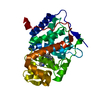
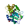
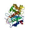
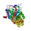
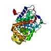
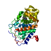
| Title | Engineering Calcium-binding site into Cytochrome c Peroxidase (CcP) | ||||||
 Components Components | Cytochrome c Peroxidase | ||||||
 Keywords Keywords | OXIDOREDUCTASE / Cation-binding loop / Calcium selectivity / Trp191 cationic-radical / open/closed conformer / bidentate bond / pentagonal bipyrimidal geometry / lignin peroxidase | ||||||
| Function / homology |  Function and homology informationcytochrome-c peroxidase / cytochrome-c peroxidase activity / response to reactive oxygen species / hydrogen peroxide catabolic process / peroxidase activity / mitochondrial intermembrane space / cellular response to oxidative stress / mitochondrial matrix / heme binding / mitochondrion / metal ion binding Function and homology informationcytochrome-c peroxidase / cytochrome-c peroxidase activity / response to reactive oxygen species / hydrogen peroxide catabolic process / peroxidase activity / mitochondrial intermembrane space / cellular response to oxidative stress / mitochondrial matrix / heme binding / mitochondrion / metal ion bindingSimilarity search - Function | ||||||
| Biological species |  Saccharomyces cerevisiae (brewer's yeast) Saccharomyces cerevisiae (brewer's yeast) | ||||||
| Method | X-RAY DIFFRACTION / MOLECULAR REPLACEMENT / Resolution: 2 Å | ||||||
 Authors Authors | Bonagura, C.A. / Bhaskar, B. / Sundaramoorthy, M. / Poulos, T.L. | ||||||
 Citation Citation | Journal: J.Biol.Chem. / Year: 1999 Title: Conversion of an engineered potassium-binding site into a calcium-selective site in cytochrome c peroxidase. Authors: Bonagura, C.A. / Bhaskar, B. / Sundaramoorthy, M. / Poulos, T.L. | ||||||
| History |
| ||||||
| Remark 400 | COMPOUND Largely a helical bundle protein containing a distinct distal and proximal heme domains. ...COMPOUND Largely a helical bundle protein containing a distinct distal and proximal heme domains. Proximal heme domain contains the cation-binding loop which in the present case contains Calcium coordinated by the residues 190-199 of the loop. |
- Structure visualization
| Structure viewer | Molecule: MolmilJmol/JSmol |
|---|
- Downloads & links
-Download
| PDBx/mmCIF format | 1krj.cif.gz | 88.9 KB | Display | PDBx/mmCIF format |
|---|---|---|---|---|
| PDB format | pdb1krj.ent.gz | 66.6 KB | Display | PDB format |
| PDBx/mmJSON format | 1krj.json.gz | Tree view | PDBx/mmJSON format | |
| Others |  Other downloads Other downloads |
-Validation report
| Arichive directory | https://data.pdbj.org/pub/pdb/validation_reports/kr/1krjftp://data.pdbj.org/pub/pdb/validation_reports/kr/1krj | HTTPS FTP |
|---|
-Related structure data
| Similar structure data |
|---|


-Links
 PDBj
PDBj
- Assembly
| Deposited unit | 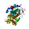
| ||||||||
|---|---|---|---|---|---|---|---|---|---|
| 1 |
| ||||||||
| Unit cell |
| ||||||||
| Details | Largely a helical bundle protein containing a distinct distal and proximal heme domains. Proximal heme domian contains the cation-binding loop which in the present case contains Calcium coordinated by the residues 190-199 of the loop |
-Components
| #1: Protein | / CCP Mass: 33661.277 Da / Num. of mol.: 1 / Fragment: Proximal Cation-binding Loop / Mutation: A176T, G192D, A194T, T199D, E201S Source method: isolated from a genetically manipulated source Source: (gene. exp.) Saccharomyces cerevisiae (brewer's yeast)Gene: OPBYC / Plasmid: pT7CcP / Species (production host): Escherichia coli / Production host:  Escherichia coli BL21(DE3) (bacteria) / Strain (production host): BL21(DE3) / References: UniProt: P00431, cytochrome-c peroxidase Escherichia coli BL21(DE3) (bacteria) / Strain (production host): BL21(DE3) / References: UniProt: P00431, cytochrome-c peroxidase |
|---|---|
| #2: Chemical | ChemComp-K /   Mass: 39.098 Da / Num. of mol.: 1 / Source method: obtained synthetically / Formula: K Mass: 39.098 Da / Num. of mol.: 1 / Source method: obtained synthetically / Formula: K |
| #3: Chemical | ChemComp-HEM / Heme B  Mass: 616.487 Da / Num. of mol.: 1 / Source method: obtained synthetically / Formula: C34H32FeN4O4 Mass: 616.487 Da / Num. of mol.: 1 / Source method: obtained synthetically / Formula: C34H32FeN4O4 |
| #4: Water | ChemComp-HOH / Water Mass: 18.015 Da / Num. of mol.: 599 / Source method: isolated from a natural source / Formula: H2O Mass: 18.015 Da / Num. of mol.: 599 / Source method: isolated from a natural source / Formula: H2O |
-Experimental details
-Experiment
| Experiment | Method: X-RAY DIFFRACTION / Number of used crystals: 1 |
|---|
- Sample preparation
| Crystal | Density Matthews: 3.06 Å3/Da / Density % sol: 59.85 % | ||||||||||||||||||||||||||||||||||||||||||
|---|---|---|---|---|---|---|---|---|---|---|---|---|---|---|---|---|---|---|---|---|---|---|---|---|---|---|---|---|---|---|---|---|---|---|---|---|---|---|---|---|---|---|---|
| Crystal grow | Temperature: 277 K / Method: vapor diffusion, sitting drop / pH: 6 Details: 2-Methyl-2,4-Pentanediol (MPD), potassium phosphate, pH 6.0, VAPOR DIFFUSION, SITTING DROP, temperature 277K | ||||||||||||||||||||||||||||||||||||||||||
| Crystal grow | *PLUS Method: unknown | ||||||||||||||||||||||||||||||||||||||||||
| Components of the solutions | *PLUS
|
-Data collection
| Diffraction | Mean temperature: 110 K |
|---|---|
| Diffraction source | Source: ROTATING ANODE / Type: RIGAKU / Wavelength: 1.5418 Å |
| Detector | Type: RIGAKU / Detector: IMAGE PLATE / Date: Oct 29, 1998 / Details: Mirrors |
| Radiation | Monochromator: Graphite / Protocol: SINGLE WAVELENGTH / Monochromatic (M) / Laue (L): M / Scattering type: x-ray |
| Radiation wavelength | Wavelength: 1.5418 Å / Relative weight: 1 |
| Reflection | Resolution: 2→20 Å / Num. obs: 28764 / Observed criterion σ(F): 2 / Observed criterion σ(I): 2 / Redundancy: 10.8627 % / Rmerge(I) obs: 0.082 / Rsym value: 0.057 / Net I/σ(I): 19.2082 |
| Reflection shell | Resolution: 2→2.02 Å / Rmerge(I) obs: 0.42 / Mean I/σ(I) obs: 3.3023 / Num. unique all: 706 / Rsym value: 0.354 / % possible all: 99.3 |
| Reflection | *PLUS Lowest resolution: 20 Å / Num. obs: 28701 / % possible obs: 99.8 % / Num. measured all: 155672 |
| Reflection shell | *PLUS % possible obs: 99 % |
- Processing
| Software |
| ||||||||||||||||||||||||||||||||||||
|---|---|---|---|---|---|---|---|---|---|---|---|---|---|---|---|---|---|---|---|---|---|---|---|---|---|---|---|---|---|---|---|---|---|---|---|---|---|
| Refinement | Method to determine structure: MOLECULAR REPLACEMENT Starting model: Potassium-binding CcP (CcPK2) Resolution: 2→10 Å / Isotropic thermal model: Isotropic / Cross valid method: THROUGHOUT / σ(F): 2 / σ(I): 2 / Stereochemistry target values: Engh & Huber
| ||||||||||||||||||||||||||||||||||||
| Refinement step | Cycle: LAST / Resolution: 2→10 Å
| ||||||||||||||||||||||||||||||||||||
| Refine LS restraints |
| ||||||||||||||||||||||||||||||||||||
| LS refinement shell |
| ||||||||||||||||||||||||||||||||||||
| Software | *PLUS Name: X-PLOR / Classification: refinement | ||||||||||||||||||||||||||||||||||||
| Refinement | *PLUS Lowest resolution: 10 Å / σ(F): 2 / Rfactor obs: 0.192 | ||||||||||||||||||||||||||||||||||||
| Solvent computation | *PLUS | ||||||||||||||||||||||||||||||||||||
| Displacement parameters | *PLUS | ||||||||||||||||||||||||||||||||||||
| Refine LS restraints | *PLUS
|
