 Movie
Movie Controller


+ Open data

- Basic information
| Entry | Database: PDB / ID: 1kqx | ||||||
|---|---|---|---|---|---|---|---|
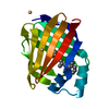
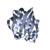
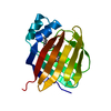
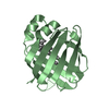
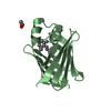
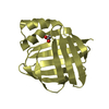
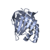
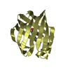
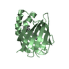
| Title | Crystal structure of apo-CRBP from zebrafish | ||||||
 Components Components | Cellular retinol-binding protein | ||||||
 Keywords Keywords |  TRANSPORT PROTEIN / retinol / vitamin A / retinol-binding TRANSPORT PROTEIN / retinol / vitamin A / retinol-binding | ||||||
| Function / homology |  Function and homology information Function and homology informationRetinoid metabolism and transport / fatty acid transport / fatty acid binding / nucleus / cytosolSimilarity search - Function | ||||||
| Biological species |  Danio rerio (zebrafish) Danio rerio (zebrafish) | ||||||
| Method | X-RAY DIFFRACTION / SYNCHROTRON / MOLECULAR REPLACEMENT / Resolution: 1.7 Å | ||||||
 Authors Authors | Calderone, V. / Folli, C. / Marchesani, A. / Berni, R. / Zanotti, G. | ||||||
 Citation Citation | Journal: J.Mol.Biol. / Year: 2002 Title: Identification and structural analysis of a zebrafish apo and holo cellular retinol-binding protein. Authors: Calderone, V. / Folli, C. / Marchesani, A. / Berni, R. / Zanotti, G. #1: Journal: J.Mol.Biol. / Year: 1993Title: Crystallographic Studies on a Family of Cellular Lipophilic Transport Proteins. Refinement of P2 Myelin Protein and the Structure Determination and Refinement of Cellular Retinol-binding ...Title: Crystallographic Studies on a Family of Cellular Lipophilic Transport Proteins. Refinement of P2 Myelin Protein and the Structure Determination and Refinement of Cellular Retinol-binding Protein in Complex with All-trans-retinol Authors: Cowan, S.W. / Newcomer, M.E. / Jones, T.A. #2: Journal: J.Mol.Biol. / Year: 1993Title: Crystal structures of holo and apo-cellular retinol-binding protein II Authors: Winter, N.S. / Bratt, J.M. / Banaszak, L.J. #3: Journal: Proc.Natl.Acad.Sci.USA / Year: 2001Title: Identification, retinoid binding and X-ray analysis of a human retinol-binding protein Authors: Folli, C. / Calderone, V. / Ottonello, S. / Bolchi, A. / Zanotti, G. / Stoppini, M. / Berni, R. | ||||||
| History |
|
- Structure visualization
| Structure viewer | Molecule: MolmilJmol/JSmol |
|---|
- Downloads & links
-Download
| PDBx/mmCIF format | 1kqx.cif.gz | 42.6 KB | Display | PDBx/mmCIF format |
|---|---|---|---|---|
| PDB format | pdb1kqx.ent.gz | 29.6 KB | Display | PDB format |
| PDBx/mmJSON format | 1kqx.json.gz | Tree view | PDBx/mmJSON format | |
| Others |  Other downloads Other downloads |
-Validation report
| Arichive directory | https://data.pdbj.org/pub/pdb/validation_reports/kq/1kqxftp://data.pdbj.org/pub/pdb/validation_reports/kq/1kqx | HTTPS FTP |
|---|
-Related structure data


-Links
 PDBj
PDBj

- Assembly
| Deposited unit | 
| ||||||||
|---|---|---|---|---|---|---|---|---|---|
| 1 |
| ||||||||
| Unit cell |
|
-Components
| #1: Protein | Mass: 15699.820 Da / Num. of mol.: 1 Source method: isolated from a genetically manipulated source Source: (gene. exp.) Danio rerio (zebrafish) / Production host:  Escherichia coli (E. coli) / References: UniProt: Q8UVG6 Escherichia coli (E. coli) / References: UniProt: Q8UVG6 |
|---|---|
| #2: Water | ChemComp-HOH / Water Mass: 18.015 Da / Num. of mol.: 126 / Source method: isolated from a natural source / Formula: H2O Mass: 18.015 Da / Num. of mol.: 126 / Source method: isolated from a natural source / Formula: H2O |
-Experimental details
-Experiment
| Experiment | Method: X-RAY DIFFRACTION / Number of used crystals: 1 |
|---|
- Sample preparation
| Crystal | Density Matthews: 2.09 Å3/Da / Density % sol: 48.98 % | ||||||||||||||||||||||||||||||
|---|---|---|---|---|---|---|---|---|---|---|---|---|---|---|---|---|---|---|---|---|---|---|---|---|---|---|---|---|---|---|---|
| Crystal grow | Temperature: 293 K / Method: vapor diffusion, sitting drop / pH: 5.6 Details: PEG 4000, Ammonium acetate, sodium citrate, pH 5.6, VAPOR DIFFUSION, SITTING DROP, temperature 293K | ||||||||||||||||||||||||||||||
| Crystal grow | *PLUS Temperature: 4 ℃ | ||||||||||||||||||||||||||||||
| Components of the solutions | *PLUS
|
-Data collection
| Diffraction | Mean temperature: 100 K |
|---|---|
| Diffraction source | Source: SYNCHROTRON / Site: ESRF  / Beamline: ID14-4 / Wavelength: 0.9393 Å / Beamline: ID14-4 / Wavelength: 0.9393 Å |
| Detector | Type: ADSC QUANTUM 4 / Detector: CCD / Date: Sep 15, 2001 |
| Radiation | Protocol: SINGLE WAVELENGTH / Monochromatic (M) / Laue (L): M / Scattering type: x-ray |
| Radiation wavelength | Wavelength: 0.9393 Å / Relative weight: 1 |
| Reflection | Resolution: 1.7→40 Å / Num. all: 16380 / Num. obs: 16380 / % possible obs: 97.7 % / Observed criterion σ(F): 0 / Observed criterion σ(I): 0 / Redundancy: 3.4 % / Rmerge(I) obs: 0.094 / Rsym value: 0.094 / Net I/σ(I): 4.6 |
| Reflection shell | Resolution: 1.7→1.79 Å / Redundancy: 2.8 % / Rmerge(I) obs: 0.294 / Mean I/σ(I) obs: 2.1 / Num. unique all: 2191 / Rsym value: 0.294 / % possible all: 92 |
| Reflection | *PLUS Highest resolution: 1.7 Å / Lowest resolution: 28 Å / Num. obs: 16261 / Num. measured all: 143455 / Rmerge(I) obs: 0.094 |
| Reflection shell | *PLUS % possible obs: 97.7 % / Rmerge(I) obs: 0.294 |
- Processing
| Software |
| |||||||||||||||||||||||||||||||||
|---|---|---|---|---|---|---|---|---|---|---|---|---|---|---|---|---|---|---|---|---|---|---|---|---|---|---|---|---|---|---|---|---|---|---|
| Refinement | Method to determine structure: MOLECULAR REPLACEMENT Starting model: holo rat CRBPII Resolution: 1.7→28.06 Å / Num. parameters: 4963 / Num. restraintsaints: 4485 / Isotropic thermal model: isotropic / Cross valid method: FREE R / σ(F): 0 / σ(I): 0 / Stereochemistry target values: ENGH AND HUBER
| |||||||||||||||||||||||||||||||||
| Solvent computation | Solvent model: MOEWS & KRETSINGER, J.MOL.BIOL.91(1973)201-228 | |||||||||||||||||||||||||||||||||
| Refine analyze | Num. disordered residues: 0 / Occupancy sum hydrogen: 0 / Occupancy sum non hydrogen: 1237 | |||||||||||||||||||||||||||||||||
| Refinement step | Cycle: LAST / Resolution: 1.7→28.06 Å
| |||||||||||||||||||||||||||||||||
| Refine LS restraints |
| |||||||||||||||||||||||||||||||||
| Software | *PLUS Name: SHELXL / Version: 97 / Classification: refinement | |||||||||||||||||||||||||||||||||
| Refinement | *PLUS Rfactor all: 0.2032 / Rfactor Rfree: 0.276 / Rfactor Rwork: 0.187 | |||||||||||||||||||||||||||||||||
| Solvent computation | *PLUS | |||||||||||||||||||||||||||||||||
| Displacement parameters | *PLUS | |||||||||||||||||||||||||||||||||
| Refine LS restraints | *PLUS
| |||||||||||||||||||||||||||||||||
| LS refinement shell | *PLUS Rfactor Rwork: 0.236 |
