 Movie
Movie Controller


+ Open data

- Basic information
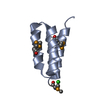
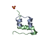
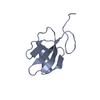
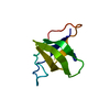
| Entry | Database: PDB / ID: 1koy | ||||||
|---|---|---|---|---|---|---|---|
| Title | NMR structure of DFF-C domain | ||||||
 Components Components | DNA fragmentation factor alpha subunit | ||||||
 Keywords Keywords |  APOPTOSIS / DFF / RIKEN Structural Genomics/Proteomics Initiative / RSGI / Structural Genomics APOPTOSIS / DFF / RIKEN Structural Genomics/Proteomics Initiative / RSGI / Structural Genomics | ||||||
| Function / homology |  Function and homology information Function and homology informationnegative regulation of deoxyribonuclease activity / deoxyribonuclease inhibitor activity / negative regulation of apoptotic DNA fragmentation / apoptotic DNA fragmentation / negative regulation of execution phase of apoptosis / Apoptosis induced DNA fragmentation / thymocyte apoptotic process / chaperone-mediated protein folding / protein folding chaperone / positive regulation of apoptotic process ...negative regulation of deoxyribonuclease activity / deoxyribonuclease inhibitor activity / negative regulation of apoptotic DNA fragmentation / apoptotic DNA fragmentation / negative regulation of execution phase of apoptosis / Apoptosis induced DNA fragmentation / thymocyte apoptotic process / chaperone-mediated protein folding / protein folding chaperone / positive regulation of apoptotic process / protein domain specific binding / chromatin / protein-containing complex / nucleoplasm / nucleus / plasma membrane / cytosolSimilarity search - Function | ||||||
| Biological species |  Homo sapiens (human) Homo sapiens (human) | ||||||
| Method | SOLUTION NMR / simulated annealing | ||||||
 Authors Authors | Fukushima, K. / Kikuchi, J. / Koshiba, S. / Kigawa, T. / Kuroda, Y. / Yokoyama, S. / RIKEN Structural Genomics/Proteomics Initiative (RSGI) | ||||||
 Citation Citation | Journal: J.Mol.Biol. / Year: 2002 Title: Solution structure of the DFF-C domain of DFF45/ICAD. A structural basis for the regulation of apoptotic DNA fragmentation. Authors: Fukushima, K. / Kikuchi, J. / Koshiba, S. / Kigawa, T. / Kuroda, Y. / Yokoyama, S. | ||||||
| History |
|
- Structure visualization
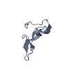
| Structure viewer | Molecule: MolmilJmol/JSmol |
|---|
- Downloads & links
-Download
| PDBx/mmCIF format | 1koy.cif.gz | 28.6 KB | Display | PDBx/mmCIF format |
|---|---|---|---|---|
| PDB format | pdb1koy.ent.gz | 19.2 KB | Display | PDB format |
| PDBx/mmJSON format | 1koy.json.gz | Tree view | PDBx/mmJSON format | |
| Others |  Other downloads Other downloads |
-Validation report
| Arichive directory | https://data.pdbj.org/pub/pdb/validation_reports/ko/1koyftp://data.pdbj.org/pub/pdb/validation_reports/ko/1koy | HTTPS FTP |
|---|
-Related structure data
| Related structure data |  1iyrC C: citing same article ( |
|---|---|
| Similar structure data | |
| Other databases |
|


-Links
 PDBj
PDBj

- Assembly
| Deposited unit | 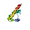
| |||||||||
|---|---|---|---|---|---|---|---|---|---|---|
| 1 |
| |||||||||
| NMR ensembles |
|
-Components
| #1: Protein | Mass: 7009.962 Da / Num. of mol.: 1 / Fragment: C-TERMINAL DOMAIN Source method: isolated from a genetically manipulated source Source: (gene. exp.) Homo sapiens (human) / Production host:  Escherichia coli (E. coli) / References: UniProt: O00273 Escherichia coli (E. coli) / References: UniProt: O00273 |
|---|
-Experimental details
-Experiment
| Experiment | Method: SOLUTION NMR |
|---|
- Sample preparation
| Details | Contents: U-13C, 15N |
|---|---|
| Sample conditions | pH: 6.50 / Temperature: 298.00 K |
| Crystal grow | *PLUS Method: other / Details: NMR |
-NMR measurement
| Radiation | Protocol: SINGLE WAVELENGTH / Monochromatic (M) / Laue (L): M |
|---|---|
| Radiation wavelength | Relative weight: 1 |
| NMR spectrometer | Type: Bruker DRX / Manufacturer: Bruker / Model: DRX / Field strength: 600 MHz |
- Processing
| NMR software |
| |||||||||
|---|---|---|---|---|---|---|---|---|---|---|
| Refinement | Method: simulated annealing / Software ordinal: 1 | |||||||||
| NMR ensemble | Conformer selection criteria: structures with the lowest energy Conformers calculated total number: 10 / Conformers submitted total number: 1 |
