 Movie
Movie Controller

[English] 日本語
 Yorodumi
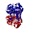
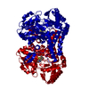
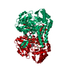
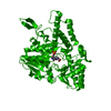
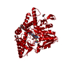
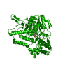
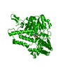
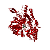
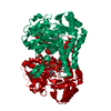
- PDB-1kmk: E. coli NifS/CsdB protein at 2.20A with the cysteine perselenide ... -
+ Open data

- Basic information
| Entry | Database: PDB / ID: 1kmk | ||||||
|---|---|---|---|---|---|---|---|
| Title | E. coli NifS/CsdB protein at 2.20A with the cysteine perselenide intermediate (residue CSZ). | ||||||
 Components Components | SELENOCYSTEINE LYASE | ||||||
 Keywords Keywords | LYASE / nifs selenocysteine cysteine persulfide perselenide xray / Structural Genomics / PSI / Protein Structure Initiative / New York SGX Research Center for Structural Genomics / NYSGXRC | ||||||
| Function / homology |  Function and homology information Function and homology informationselenium compound metabolic process / Hydrolases; Acting on carbon-sulfur bonds; Acting on carbon-sulfur bonds / cysteine sulfinate desulfinase activity / selenocysteine lyase / selenocysteine lyase activity / cysteine desulfurase / cysteine desulfurase activity / sulfur compound metabolic process / cysteine metabolic process / iron-sulfur cluster assembly ...selenium compound metabolic process / Hydrolases; Acting on carbon-sulfur bonds; Acting on carbon-sulfur bonds / cysteine sulfinate desulfinase activity / selenocysteine lyase / selenocysteine lyase activity / cysteine desulfurase / cysteine desulfurase activity / sulfur compound metabolic process / cysteine metabolic process / iron-sulfur cluster assembly / pyridoxal phosphate binding / hydrolase activity / protein homodimerization activity / cytoplasmSimilarity search - Function | ||||||
| Biological species |  Escherichia coli (E. coli) Escherichia coli (E. coli) | ||||||
| Method | X-RAY DIFFRACTION / SYNCHROTRON / starting model 1jf9 / Resolution: 2.2 Å | ||||||
 Authors Authors | Lima, C.D. / Burley, S.K. / New York SGX Research Center for Structural Genomics (NYSGXRC) | ||||||
 Citation Citation | Journal: J.Mol.Biol. / Year: 2002 Title: Analysis of the E. coli NifS CsdB protein at 2.0 A reveals the structural basis for perselenide and persulfide intermediate formation. Authors: Lima, C.D. | ||||||
| History |
|
- Structure visualization
| Structure viewer | Molecule: MolmilJmol/JSmol |
|---|
- Downloads & links
-Download
| PDBx/mmCIF format | 1kmk.cif.gz | 104.5 KB | Display | PDBx/mmCIF format |
|---|---|---|---|---|
| PDB format | pdb1kmk.ent.gz | 78.3 KB | Display | PDB format |
| PDBx/mmJSON format | 1kmk.json.gz | Tree view | PDBx/mmJSON format | |
| Others |  Other downloads Other downloads |
-Validation report
| Arichive directory | https://data.pdbj.org/pub/pdb/validation_reports/km/1kmkftp://data.pdbj.org/pub/pdb/validation_reports/km/1kmk | HTTPS FTP |
|---|
-Related structure data
| Related structure data |  1jf9SC  1kmjC S: Starting model for refinement C: citing same article ( |
|---|---|
| Similar structure data | |
| Other databases |


-Links
 PDBj
PDBj- Assembly
| Deposited unit | 
| ||||||||
|---|---|---|---|---|---|---|---|---|---|
| 1 | 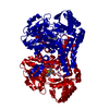
| ||||||||
| Unit cell |
|
-Components
| #1: Protein | / NIFS/CSDB / SELENOCYSTEINE REDUCTASE / SELENOCYSTEINE BETA-LYASE / SCL Mass: 44559.555 Da / Num. of mol.: 1 Source method: isolated from a genetically manipulated source Source: (gene. exp.) Escherichia coli (E. coli) / Production host: Escherichia coli (E. coli) / References: UniProt: P77444, selenocysteine lyase |
|---|---|
| #2: Chemical | ChemComp-SEC / Selenocysteine  Type: L-peptide linking / Mass: 168.053 Da / Num. of mol.: 1 / Source method: obtained synthetically / Formula: C3H7NO2Se Type: L-peptide linking / Mass: 168.053 Da / Num. of mol.: 1 / Source method: obtained synthetically / Formula: C3H7NO2Se |
| #3: Chemical | ChemComp-PLP / Pyridoxal phosphate  Mass: 247.142 Da / Num. of mol.: 1 / Source method: obtained synthetically / Formula: C8H10NO6P Mass: 247.142 Da / Num. of mol.: 1 / Source method: obtained synthetically / Formula: C8H10NO6P |
| #4: Water | ChemComp-HOH / Water Mass: 18.015 Da / Num. of mol.: 485 / Source method: isolated from a natural source / Formula: H2O Mass: 18.015 Da / Num. of mol.: 485 / Source method: isolated from a natural source / Formula: H2O |
-Experimental details
-Experiment
| Experiment | Method: X-RAY DIFFRACTION / Number of used crystals: 1 |
|---|
- Sample preparation
| Crystal grow | Temperature: 291 K / Method: vapor diffusion, hanging drop / pH: 7 Details: 4M NACL, pH 7, VAPOR DIFFUSION, HANGING DROP, temperature 291K | |||||||||||||||||||||||||||||||||||||||||||||||||
|---|---|---|---|---|---|---|---|---|---|---|---|---|---|---|---|---|---|---|---|---|---|---|---|---|---|---|---|---|---|---|---|---|---|---|---|---|---|---|---|---|---|---|---|---|---|---|---|---|---|---|
| Crystal grow | *PLUS pH: 8 | |||||||||||||||||||||||||||||||||||||||||||||||||
| Components of the solutions | *PLUS
|
-Data collection
| Diffraction | Mean temperature: 100 K |
|---|---|
| Diffraction source | Source: SYNCHROTRON / Site: NSLS  / Beamline: X4A / Wavelength: 0.979 Å / Beamline: X4A / Wavelength: 0.979 Å |
| Detector | Type: ADSC QUANTUM 4 / Detector: CCD / Date: Oct 1, 2001 |
| Radiation | Monochromator: SAGITALLY FOCUSED / Protocol: SINGLE WAVELENGTH / Monochromatic (M) / Laue (L): M / Scattering type: x-ray |
| Radiation wavelength | Wavelength: 0.979 Å / Relative weight: 1 |
| Reflection | Resolution: 2.2→20 Å / Num. all: 49125 / Num. obs: 49125 / % possible obs: 87.9 % / Observed criterion σ(F): 0 / Observed criterion σ(I): 0 / Rsym value: 0.055 / Net I/σ(I): 7.2 |
| Reflection shell | Resolution: 2.2→2.28 Å / Mean I/σ(I) obs: 2.1 / Rsym value: 0.4 / % possible all: 67.3 |
| Reflection | *PLUS Num. measured all: 1806159 / Rmerge(I) obs: 0.055 |
| Reflection shell | *PLUS % possible obs: 67.3 % / Rmerge(I) obs: 0.4 |
- Processing
| Software |
| |||||||||||||||||||||||||
|---|---|---|---|---|---|---|---|---|---|---|---|---|---|---|---|---|---|---|---|---|---|---|---|---|---|---|
| Refinement | Method to determine structure: starting model 1jf9 Starting model: 1jf9 Resolution: 2.2→20 Å / Cross valid method: Rfree test / σ(F): 0 / σ(I): 0 / Stereochemistry target values: ENGH AND HUBER, 1991
| |||||||||||||||||||||||||
| Refinement step | Cycle: LAST / Resolution: 2.2→20 Å
| |||||||||||||||||||||||||
| Refine LS restraints |
| |||||||||||||||||||||||||
| Refinement | *PLUS % reflection Rfree: 5 % / Rfactor obs: 0.196 | |||||||||||||||||||||||||
| Solvent computation | *PLUS | |||||||||||||||||||||||||
| Displacement parameters | *PLUS |
