 Movie
Movie Controller


+ Open data

- Basic information
| Entry | Database: PDB / ID: 1kiz | ||||||
|---|---|---|---|---|---|---|---|
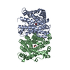
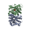
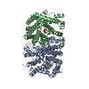
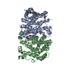
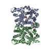
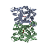
| Title | D100E trichodiene synthase complexed with pyrophosphate | ||||||
 Components Components | trichodiene synthase | ||||||
 Keywords Keywords | LYASE / terpenoid synthase fold / site-directed mutant / pyrophosphate | ||||||
| Function / homology |  Function and homology informationtrichodiene synthase / sesquiterpenoid biosynthetic process / trichodiene synthase activity / metal ion binding Function and homology informationtrichodiene synthase / sesquiterpenoid biosynthetic process / trichodiene synthase activity / metal ion bindingSimilarity search - Function | ||||||
| Biological species |  Fusarium sporotrichioides (fungus) Fusarium sporotrichioides (fungus) | ||||||
| Method | X-RAY DIFFRACTION / SYNCHROTRON / FOURIER SYNTHESIS / Resolution: 2.6 Å | ||||||
 Authors Authors | Rynkiewicz, M.J. / Cane, D.E. / Christianson, D.W. | ||||||
 Citation Citation | Journal: Biochemistry / Year: 2002 Title: X-ray crystal structures of D100E trichodiene synthase and its pyrophosphate complex reveal the basis for terpene product diversity. Authors: Rynkiewicz, M.J. / Cane, D.E. / Christianson, D.W. | ||||||
| History |
|
- Structure visualization
| Structure viewer | Molecule: MolmilJmol/JSmol |
|---|
- Downloads & links
-Download
| PDBx/mmCIF format | 1kiz.cif.gz | 160.9 KB | Display | PDBx/mmCIF format |
|---|---|---|---|---|
| PDB format | pdb1kiz.ent.gz | 127.5 KB | Display | PDB format |
| PDBx/mmJSON format | 1kiz.json.gz | Tree view | PDBx/mmJSON format | |
| Others |  Other downloads Other downloads |
-Validation report
| Arichive directory | https://data.pdbj.org/pub/pdb/validation_reports/ki/1kizftp://data.pdbj.org/pub/pdb/validation_reports/ki/1kiz | HTTPS FTP |
|---|
-Related structure data
| Related structure data |  1kiyC 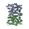 1jfgS S: Starting model for refinement C: citing same article ( |
|---|---|
| Similar structure data |


-Links
 PDBj
PDBj


- Assembly
| Deposited unit | 
| ||||||||
|---|---|---|---|---|---|---|---|---|---|
| 1 |
| ||||||||
| Unit cell |
| ||||||||
| Details | To generate the biological dimer from the asymmetric unit, apply tranformation 6_665 to chain A and 6_655 to chain B |
-Components
| #1: Protein | Mass: 44064.629 Da / Num. of mol.: 2 / Mutation: D100E Source method: isolated from a genetically manipulated source Source: (gene. exp.) Fusarium sporotrichioides (fungus) / Gene: Tri5 / Species (production host): Escherichia coli / Production host:  Escherichia coli BL21(DE3) (bacteria) / Strain (production host): BL21(DE3) / References: UniProt: P13513, EC: 4.1.99.6 Escherichia coli BL21(DE3) (bacteria) / Strain (production host): BL21(DE3) / References: UniProt: P13513, EC: 4.1.99.6#2: Chemical | ChemComp-EDO / Ethylene glycol  Mass: 62.068 Da / Num. of mol.: 4 / Source method: obtained synthetically / Formula: C2H6O2 Mass: 62.068 Da / Num. of mol.: 4 / Source method: obtained synthetically / Formula: C2H6O2#3: Chemical |   Mass: 24.305 Da / Num. of mol.: 2 / Source method: obtained synthetically / Formula: Mg Mass: 24.305 Da / Num. of mol.: 2 / Source method: obtained synthetically / Formula: Mg#4: Chemical | ChemComp-POP / | Pyrophosphate  Mass: 175.959 Da / Num. of mol.: 1 / Source method: obtained synthetically / Formula: H2O7P2 Mass: 175.959 Da / Num. of mol.: 1 / Source method: obtained synthetically / Formula: H2O7P2#5: Water | ChemComp-HOH / | Water Mass: 18.015 Da / Num. of mol.: 226 / Source method: isolated from a natural source / Formula: H2O Mass: 18.015 Da / Num. of mol.: 226 / Source method: isolated from a natural source / Formula: H2O |
|---|
-Experimental details
-Experiment
| Experiment | Method: X-RAY DIFFRACTION / Number of used crystals: 1 |
|---|
- Sample preparation
| Crystal | Density Matthews: 3.72 Å3/Da / Density % sol: 66.97 % | |||||||||||||||||||||||||||||||||||
|---|---|---|---|---|---|---|---|---|---|---|---|---|---|---|---|---|---|---|---|---|---|---|---|---|---|---|---|---|---|---|---|---|---|---|---|---|
| Crystal grow | Temperature: 277 K / Method: vapor diffusion, hanging drop / pH: 6.9 Details: PEG 8000, calcium chloride, sodium HEPES, pH 6.9, VAPOR DIFFUSION, HANGING DROP, temperature 277K | |||||||||||||||||||||||||||||||||||
| Crystal grow | *PLUS Details: Rynkiewicz, M.J., (2001) Proc.Natl.Acad.Sci.U.S.J., 98, 13543. | |||||||||||||||||||||||||||||||||||
| Components of the solutions | *PLUS
|
-Data collection
| Diffraction | Mean temperature: 100 K |
|---|---|
| Diffraction source | Source: SYNCHROTRON / Site: APS  / Beamline: 19-ID / Wavelength: 1 Å / Beamline: 19-ID / Wavelength: 1 Å |
| Detector | Type: SBC-2 / Detector: CCD / Date: Feb 14, 2001 |
| Radiation | Monochromator: SI-111 + SI-220 / Protocol: SINGLE WAVELENGTH / Monochromatic (M) / Laue (L): M / Scattering type: x-ray |
| Radiation wavelength | Wavelength: 1 Å / Relative weight: 1 |
| Reflection | Resolution: 2.6→25 Å / Num. all: 41296 / Num. obs: 41214 / % possible obs: 99.8 % / Observed criterion σ(I): -3 / Redundancy: 6 % / Biso Wilson estimate: 42.2 Å2 |
| Reflection shell | Resolution: 2.59→2.69 Å / Redundancy: 6.2 % / % possible all: 100 |
| Reflection | *PLUS Lowest resolution: 25 Å / Num. measured all: 246608 / Rmerge(I) obs: 0.081 |
| Reflection shell | *PLUS % possible obs: 100 % / Rmerge(I) obs: 0.237 |
- Processing
| Software |
| ||||||||||||||||||||||||
|---|---|---|---|---|---|---|---|---|---|---|---|---|---|---|---|---|---|---|---|---|---|---|---|---|---|
| Refinement | Method to determine structure: FOURIER SYNTHESIS Starting model: PDB CODE 1JFG Resolution: 2.6→24.49 Å / Rfactor Rfree error: 0.005 / Data cutoff high absF: 2061956.09 / Data cutoff low absF: 0 / Isotropic thermal model: RESTRAINED / Cross valid method: THROUGHOUT / σ(F): 0 / Stereochemistry target values: Engh & Huber
| ||||||||||||||||||||||||
| Solvent computation | Solvent model: FLAT MODEL / Bsol: 31.3452 Å2 / ksol: 0.324764 e/Å3 | ||||||||||||||||||||||||
| Displacement parameters | Biso mean: 44.3 Å2
| ||||||||||||||||||||||||
| Refine analyze |
| ||||||||||||||||||||||||
| Refinement step | Cycle: LAST / Resolution: 2.6→24.49 Å
| ||||||||||||||||||||||||
| Refine LS restraints |
| ||||||||||||||||||||||||
| LS refinement shell | Resolution: 2.6→2.76 Å / Rfactor Rfree error: 0.014 / Total num. of bins used: 6
| ||||||||||||||||||||||||
| Xplor file |
| ||||||||||||||||||||||||
| Refinement | *PLUS Highest resolution: 2.6 Å / Lowest resolution: 25 Å / σ(F): 0 / % reflection Rfree: 7.6 % / Rfactor obs: 0.223 / Rfactor Rfree: 0.256 | ||||||||||||||||||||||||
| Solvent computation | *PLUS | ||||||||||||||||||||||||
| Displacement parameters | *PLUS Biso mean: 44.3 Å2 | ||||||||||||||||||||||||
| Refine LS restraints | *PLUS
| ||||||||||||||||||||||||
| LS refinement shell | *PLUS Rfactor Rfree: 0.334 / % reflection Rfree: 8.5 % / Rfactor Rwork: 0.282 |
