 Movie
Movie Controller

[English] 日本語
 Yorodumi
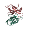
- PDB-1kgd: Crystal Structure of the Guanylate Kinase-like Domain of Human CASK -
+ Open data

- Basic information
| Entry | Database: PDB / ID: 1kgd | ||||||
|---|---|---|---|---|---|---|---|
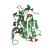
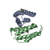
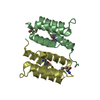
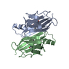
| Title | Crystal Structure of the Guanylate Kinase-like Domain of Human CASK | ||||||
 Components Components | PERIPHERAL PLASMA MEMBRANE CASK | ||||||
 Keywords Keywords |  PROTEIN BINDING / MAGUK / CASK / GUANYLATE KINASE LIKE DOMAIN PROTEIN BINDING / MAGUK / CASK / GUANYLATE KINASE LIKE DOMAIN | ||||||
| Function / homology |  Function and homology information Function and homology informationnegative regulation of cellular response to growth factor stimulus / guanylate kinase activity / Dopamine Neurotransmitter Release Cycle / neurexin family protein binding / nuclear lamina / regulation of neurotransmitter secretion / negative regulation of wound healing / Assembly and cell surface presentation of NMDA receptors / calcium ion import / Neurexins and neuroligins ...negative regulation of cellular response to growth factor stimulus / guanylate kinase activity / Dopamine Neurotransmitter Release Cycle / neurexin family protein binding / nuclear lamina / regulation of neurotransmitter secretion / negative regulation of wound healing / Assembly and cell surface presentation of NMDA receptors / calcium ion import / Neurexins and neuroligins / Sensory processing of sound by outer hair cells of the cochlea / Sensory processing of sound by inner hair cells of the cochlea / Nephrin family interactions / ciliary membrane / regulation of synaptic vesicle exocytosis / Syndecan interactions / negative regulation of cell-matrix adhesion / positive regulation of calcium ion import / basement membrane / negative regulation of keratinocyte proliferation / establishment of localization in cell / Schaffer collateral - CA1 synapse / nuclear matrix / cell-cell junction / actin cytoskeleton / presynaptic membrane / basolateral plasma membrane / vesicle / calmodulin binding / non-specific serine/threonine protein kinase / cell adhesion / phosphorylation / signaling receptor binding / focal adhesion / protein serine kinase activity / protein serine/threonine kinase activity / nucleolus / positive regulation of transcription by RNA polymerase II / ATP binding / plasma membrane / cytosol / cytoplasmSimilarity search - Function | ||||||
| Biological species |  Homo sapiens (human) Homo sapiens (human) | ||||||
| Method | X-RAY DIFFRACTION / SYNCHROTRON / MAD / Resolution: 1.314 Å | ||||||
 Authors Authors | Li, Y. / Spangenberg, O. / Paarmann, I. / Konrad, M. / Lavie, A. | ||||||
 Citation Citation | Journal: J.Biol.Chem. / Year: 2002 Title: Structural basis for nucleotide-dependent regulation of membrane-associated guanylate kinase-like domains. Authors: Li, Y. / Spangenberg, O. / Paarmann, I. / Konrad, M. / Lavie, A. | ||||||
| History |
|
- Structure visualization
| Structure viewer | Molecule: MolmilJmol/JSmol |
|---|
- Downloads & links
-Download
| PDBx/mmCIF format | 1kgd.cif.gz | 92.9 KB | Display | PDBx/mmCIF format |
|---|---|---|---|---|
| PDB format | pdb1kgd.ent.gz | 72.3 KB | Display | PDB format |
| PDBx/mmJSON format | 1kgd.json.gz | Tree view | PDBx/mmJSON format | |
| Others |  Other downloads Other downloads |
-Validation report
| Arichive directory | https://data.pdbj.org/pub/pdb/validation_reports/kg/1kgdftp://data.pdbj.org/pub/pdb/validation_reports/kg/1kgd | HTTPS FTP |
|---|
-Related structure data
| Similar structure data |
|---|


-Links
 PDBj
PDBj


- Assembly
| Deposited unit | 
| ||||||||
|---|---|---|---|---|---|---|---|---|---|
| 1 |
| ||||||||
| Unit cell |
|
-Components
| #1: Protein | Mass: 20754.535 Da / Num. of mol.: 1 / Fragment: guanylate kinase like domain Source method: isolated from a genetically manipulated source Source: (gene. exp.) Homo sapiens (human) / Production host:  Escherichia coli (E. coli) / References: UniProt: O14936 Escherichia coli (E. coli) / References: UniProt: O14936 | ||
|---|---|---|---|
| #2: Chemical | ChemComp-FMT / Formic acid  Mass: 46.025 Da / Num. of mol.: 5 / Source method: obtained synthetically / Formula: CH2O2 Mass: 46.025 Da / Num. of mol.: 5 / Source method: obtained synthetically / Formula: CH2O2#3: Water | ChemComp-HOH / | Water Mass: 18.015 Da / Num. of mol.: 218 / Source method: isolated from a natural source / Formula: H2O Mass: 18.015 Da / Num. of mol.: 218 / Source method: isolated from a natural source / Formula: H2O |
-Experimental details
-Experiment
| Experiment | Method: X-RAY DIFFRACTION / Number of used crystals: 1 |
|---|
- Sample preparation
| Crystal | Density Matthews: 2.57 Å3/Da / Density % sol: 52.23 % | ||||||||||||||||||||||||||||||||||||||||||||||||||||||||
|---|---|---|---|---|---|---|---|---|---|---|---|---|---|---|---|---|---|---|---|---|---|---|---|---|---|---|---|---|---|---|---|---|---|---|---|---|---|---|---|---|---|---|---|---|---|---|---|---|---|---|---|---|---|---|---|---|---|
| Crystal grow | Temperature: 298 K / Method: vapor diffusion, hanging drop / pH: 7.5 Details: sodium formate, potassium chloride, magnesium chloride, Tris, pH 7.5, VAPOR DIFFUSION, HANGING DROP, temperature 298.0K | ||||||||||||||||||||||||||||||||||||||||||||||||||||||||
| Crystal grow | *PLUS Temperature: 20 ℃ | ||||||||||||||||||||||||||||||||||||||||||||||||||||||||
| Components of the solutions | *PLUS
|
-Data collection
| Diffraction | Mean temperature: 100 K |
|---|---|
| Diffraction source | Source: SYNCHROTRON / Site: APS  / Beamline: 14-BM-D / Beamline: 14-BM-D |
| Detector | Type: ADSC QUANTUM 4 / Detector: CCD / Date: Mar 22, 2001 |
| Radiation | Protocol: SINGLE WAVELENGTH / Monochromatic (M) / Laue (L): M / Scattering type: x-ray |
| Radiation wavelength | Relative weight: 1 |
| Reflection | Resolution: 1.31→50 Å / Num. obs: 48853 / % possible obs: 94.2 % / Redundancy: 6.7 % / Rsym value: 0.034 / Net I/σ(I): 20.9 |
| Reflection shell | Resolution: 1.31→1.4 Å / Redundancy: 5.2 % / Mean I/σ(I) obs: 8.31 / Rsym value: 0.145 / % possible all: 81 |
| Reflection | *PLUS Num. measured all: 327011 / Rmerge(I) obs: 0.034 |
| Reflection shell | *PLUS % possible obs: 81 % / Rmerge(I) obs: 0.145 |
- Processing
| Software |
| ||||||||||||||||||||||||||||||||||||||||||||||||||||||||||||||||||||||||||||||||||||||||||||||||||||||||||||||||||||||||||||||||||
|---|---|---|---|---|---|---|---|---|---|---|---|---|---|---|---|---|---|---|---|---|---|---|---|---|---|---|---|---|---|---|---|---|---|---|---|---|---|---|---|---|---|---|---|---|---|---|---|---|---|---|---|---|---|---|---|---|---|---|---|---|---|---|---|---|---|---|---|---|---|---|---|---|---|---|---|---|---|---|---|---|---|---|---|---|---|---|---|---|---|---|---|---|---|---|---|---|---|---|---|---|---|---|---|---|---|---|---|---|---|---|---|---|---|---|---|---|---|---|---|---|---|---|---|---|---|---|---|---|---|---|---|
| Refinement | Method to determine structure: MAD / Resolution: 1.314→12 Å / Cor.coef. Fo:Fc: 0.951 / Cor.coef. Fo:Fc free: 0.94 / SU B: 2.232 / SU ML: 0.04 / Isotropic thermal model: BABINET MODEL WITH MASK / Cross valid method: THROUGHOUT / ESU R: 0.07 / ESU R Free: 0.064 / Stereochemistry target values: Engh & Huber
| ||||||||||||||||||||||||||||||||||||||||||||||||||||||||||||||||||||||||||||||||||||||||||||||||||||||||||||||||||||||||||||||||||
| Solvent computation | Ion probe radii: 0.8 Å / Shrinkage radii: 0.8 Å / VDW probe radii: 1.4 Å | ||||||||||||||||||||||||||||||||||||||||||||||||||||||||||||||||||||||||||||||||||||||||||||||||||||||||||||||||||||||||||||||||||
| Displacement parameters | Biso mean: 25.768 Å2
| ||||||||||||||||||||||||||||||||||||||||||||||||||||||||||||||||||||||||||||||||||||||||||||||||||||||||||||||||||||||||||||||||||
| Refinement step | Cycle: LAST / Resolution: 1.314→12 Å
| ||||||||||||||||||||||||||||||||||||||||||||||||||||||||||||||||||||||||||||||||||||||||||||||||||||||||||||||||||||||||||||||||||
| Refine LS restraints |
| ||||||||||||||||||||||||||||||||||||||||||||||||||||||||||||||||||||||||||||||||||||||||||||||||||||||||||||||||||||||||||||||||||
| LS refinement shell | Resolution: 1.314→1.348 Å / Total num. of bins used: 20
| ||||||||||||||||||||||||||||||||||||||||||||||||||||||||||||||||||||||||||||||||||||||||||||||||||||||||||||||||||||||||||||||||||
| Software | *PLUS Name: REFMAC / Version: 5 / Classification: refinement | ||||||||||||||||||||||||||||||||||||||||||||||||||||||||||||||||||||||||||||||||||||||||||||||||||||||||||||||||||||||||||||||||||
| Refinement | *PLUS % reflection Rfree: 10.1 % / Rfactor obs: 0.186 | ||||||||||||||||||||||||||||||||||||||||||||||||||||||||||||||||||||||||||||||||||||||||||||||||||||||||||||||||||||||||||||||||||
| Solvent computation | *PLUS | ||||||||||||||||||||||||||||||||||||||||||||||||||||||||||||||||||||||||||||||||||||||||||||||||||||||||||||||||||||||||||||||||||
| Displacement parameters | *PLUS | ||||||||||||||||||||||||||||||||||||||||||||||||||||||||||||||||||||||||||||||||||||||||||||||||||||||||||||||||||||||||||||||||||
| Refine LS restraints | *PLUS
|
