 Movie
Movie Controller

[English] 日本語
 Yorodumi
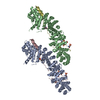
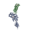
- PDB-1kfg: The X-ray Crystal Structure of Cel9G from Clostridium cellulolyti... -
+ Open data

- Basic information
| Entry | Database: PDB / ID: 1kfg | |||||||||
|---|---|---|---|---|---|---|---|---|---|---|
| Title | The X-ray Crystal Structure of Cel9G from Clostridium cellulolyticum complexed with a Thio-Oligosaccharide Inhibitor | |||||||||
 Components Components | ENDOGLUCANASE G | |||||||||
 Keywords Keywords |  HYDROLASE / endoglucanase / family 9 / thio-oligosaccharide / cellulose binding domain / (alpha-alpha)6-barrel HYDROLASE / endoglucanase / family 9 / thio-oligosaccharide / cellulose binding domain / (alpha-alpha)6-barrel | |||||||||
| Function / homology |  Function and homology informationcellulose binding / cellulase / cellulase activity / cellulose catabolic process Function and homology informationcellulose binding / cellulase / cellulase activity / cellulose catabolic processSimilarity search - Function | |||||||||
| Biological species |  Clostridium cellulolyticum (bacteria) Clostridium cellulolyticum (bacteria) | |||||||||
| Method | X-RAY DIFFRACTION / FOURIER SYNTHESIS / Resolution: 1.9 Å | |||||||||
 Authors Authors | Mandelman, D. / Belaich, A. / Belaich, J.-P. / Driguez, H. / Haser, R. | |||||||||
 Citation Citation | Journal: J.Bacteriol. / Year: 2003 Title: The X-ray crystal structure of the multidomain endoglucanase Cel9G from Clostridium cellulolyticum complexed with natural and synthetic cello-olligosaccharides Authors: Mandelman, D. / Belaich, A. / Belaich, J.-P. / Aghajari, N. / Driguez, H. / Haser, R. | |||||||||
| History |
|
- Structure visualization
| Structure viewer | Molecule: MolmilJmol/JSmol |
|---|
- Downloads & links
-Download
| PDBx/mmCIF format | 1kfg.cif.gz | 264 KB | Display | PDBx/mmCIF format |
|---|---|---|---|---|
| PDB format | pdb1kfg.ent.gz | 209.1 KB | Display | PDB format |
| PDBx/mmJSON format | 1kfg.json.gz | Tree view | PDBx/mmJSON format | |
| Others |  Other downloads Other downloads |
-Validation report
| Arichive directory | https://data.pdbj.org/pub/pdb/validation_reports/kf/1kfgftp://data.pdbj.org/pub/pdb/validation_reports/kf/1kfg | HTTPS FTP |
|---|
-Related structure data
| Related structure data |  1g87SC  1ga2C  1k72C S: Starting model for refinement C: citing same article ( |
|---|---|
| Similar structure data |


-Links
 PDBj
PDBj

- Assembly
| Deposited unit | 
| ||||||||
|---|---|---|---|---|---|---|---|---|---|
| 1 |
| ||||||||
| Unit cell |
|
-Components
-Protein / Sugars , 2 types, 4 molecules AB
| #1: Protein | Mass: 68061.086 Da / Num. of mol.: 2 / Fragment: residues 36-649 of SWS P37700 Source method: isolated from a genetically manipulated source Details: Complexed with hemithiocellodextrin GS4-GLC-GS4-GS1. Protein chain A complexed with chain X, B with Y Source: (gene. exp.) Clostridium cellulolyticum (bacteria) / Gene: CELCCG / Plasmid: pGEX-5X-2, pET-22b / Production host: Escherichia coli (E. coli) / Strain (production host): E. coli BL-21 / References: UniProt: P37700, cellulase#2: Polysaccharide | Type: oligosaccharide / Mass: 714.773 Da / Num. of mol.: 2Source method: isolated from a genetically manipulated source |
|---|
-Non-polymers , 6 types, 681 molecules 


| #3: Chemical | ChemComp-CA /  Mass: 40.078 Da / Num. of mol.: 4 / Source method: obtained synthetically / Formula: Ca Mass: 40.078 Da / Num. of mol.: 4 / Source method: obtained synthetically / Formula: Ca#4: Chemical | ChemComp-MG /  Mass: 24.305 Da / Num. of mol.: 5 / Source method: obtained synthetically / Formula: Mg Mass: 24.305 Da / Num. of mol.: 5 / Source method: obtained synthetically / Formula: Mg#5: Chemical | ChemComp-TRS / | Tris Mass: 122.143 Da / Num. of mol.: 1 / Source method: obtained synthetically / Formula: C4H12NO3 / Comment: pH buffer*YM Mass: 122.143 Da / Num. of mol.: 1 / Source method: obtained synthetically / Formula: C4H12NO3 / Comment: pH buffer*YM#6: Chemical | ChemComp-GOL / Glycerol Mass: 92.094 Da / Num. of mol.: 4 / Source method: obtained synthetically / Formula: C3H8O3 Mass: 92.094 Da / Num. of mol.: 4 / Source method: obtained synthetically / Formula: C3H8O3#7: Chemical | ChemComp-NI / | Nickel Mass: 58.693 Da / Num. of mol.: 1 / Source method: obtained synthetically / Formula: Ni Mass: 58.693 Da / Num. of mol.: 1 / Source method: obtained synthetically / Formula: Ni#8: Water | ChemComp-HOH / | WaterMass: 18.015 Da / Num. of mol.: 666 / Source method: isolated from a natural source / Formula: H2O |
|---|
-Experimental details
-Experiment
| Experiment | Method: X-RAY DIFFRACTION / Number of used crystals: 1 |
|---|
- Sample preparation
| Crystal | Density Matthews: 2.02 Å3/Da / Density % sol: 39.06 % | ||||||||||||||||||||||||||||||||||||||||||
|---|---|---|---|---|---|---|---|---|---|---|---|---|---|---|---|---|---|---|---|---|---|---|---|---|---|---|---|---|---|---|---|---|---|---|---|---|---|---|---|---|---|---|---|
| Crystal grow | Temperature: 290 K / Method: vapor diffusion, hanging drop / pH: 8.4 Details: Peg 4000, Tris, MgAcO, Isopropanol, Glycerol, pH 8.4, VAPOR DIFFUSION, HANGING DROP, temperature 290K | ||||||||||||||||||||||||||||||||||||||||||
| Crystal grow | *PLUS Temperature: 17 ℃ / Method: vapor diffusion, hanging drop | ||||||||||||||||||||||||||||||||||||||||||
| Components of the solutions | *PLUS
|
-Data collection
| Diffraction | Mean temperature: 100 K |
|---|---|
| Diffraction source | Source: ROTATING ANODE / Type: ENRAF-NONIUS FR571 / Wavelength: 1.5418 Å |
| Detector | Type: MARRESEARCH / Detector: IMAGE PLATE / Date: Mar 1, 2001 / Details: Osmic mirrors |
| Radiation | Monochromator: Osmic mirrors / Protocol: SINGLE WAVELENGTH / Monochromatic (M) / Laue (L): M / Scattering type: x-ray |
| Radiation wavelength | Wavelength: 1.5418 Å / Relative weight: 1 |
| Reflection | Resolution: 1.9→42 Å / Num. all: 95613 / Num. obs: 87964 / % possible obs: 92 % / Observed criterion σ(I): 4.8 / Redundancy: 2.7 % / Biso Wilson estimate: 15.4 Å2 / Rsym value: 0.048 / Net I/σ(I): 18.2 |
| Reflection shell | Resolution: 1.9→1.96 Å / Mean I/σ(I) obs: 4.8 / Num. unique all: 5168 / Rsym value: 0.183 / % possible all: 87.6 |
| Reflection | *PLUS Lowest resolution: 50 Å / Num. obs: 270109 / % possible obs: 88.7 % / Redundancy: 2.6 % / Rmerge(I) obs: 0.048 |
| Reflection shell | *PLUS % possible obs: 87.4 % / Rmerge(I) obs: 0.199 / Mean I/σ(I) obs: 3.8 |
- Processing
| Software |
| ||||||||||||||||||||||||||||||||||||
|---|---|---|---|---|---|---|---|---|---|---|---|---|---|---|---|---|---|---|---|---|---|---|---|---|---|---|---|---|---|---|---|---|---|---|---|---|---|
| Refinement | Method to determine structure: FOURIER SYNTHESIS Starting model: 1G87 Resolution: 1.9→42.42 Å / Rfactor Rfree error: 0.003 / Data cutoff high absF: 1381731.32 / Data cutoff low absF: 0 / Isotropic thermal model: RESTRAINED / Cross valid method: THROUGHOUT / σ(F): 0 / σ(I): 3 / Stereochemistry target values: Engh & Huber
| ||||||||||||||||||||||||||||||||||||
| Solvent computation | Solvent model: FLAT MODEL / Bsol: 50.1449 Å2 / ksol: 0.399916 e/Å3 | ||||||||||||||||||||||||||||||||||||
| Displacement parameters | Biso mean: 17.9 Å2
| ||||||||||||||||||||||||||||||||||||
| Refine analyze |
| ||||||||||||||||||||||||||||||||||||
| Refinement step | Cycle: LAST / Resolution: 1.9→42.42 Å
| ||||||||||||||||||||||||||||||||||||
| Refine LS restraints |
| ||||||||||||||||||||||||||||||||||||
| LS refinement shell | Resolution: 1.9→1.97 Å / Rfactor Rfree error: 0.013 / Total num. of bins used: 10
| ||||||||||||||||||||||||||||||||||||
| Xplor file |
| ||||||||||||||||||||||||||||||||||||
| Refinement | *PLUS Highest resolution: 1.9 Å / Lowest resolution: 50 Å | ||||||||||||||||||||||||||||||||||||
| Solvent computation | *PLUS | ||||||||||||||||||||||||||||||||||||
| Displacement parameters | *PLUS | ||||||||||||||||||||||||||||||||||||
| Refine LS restraints | *PLUS
|
