 Movie
Movie Controller


+ Open data

- Basic information
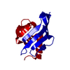
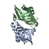
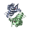
| Entry | Database: PDB / ID: 1k41 | ||||||
|---|---|---|---|---|---|---|---|
| Title | Crystal structure of KSI Y57S mutant | ||||||
 Components Components | Ketosteroid Isomerase | ||||||
 Keywords Keywords |  ISOMERASE / KSI Y57S Helix ISOMERASE / KSI Y57S Helix | ||||||
| Function / homology |  Function and homology informationsteroid Delta-isomerase / steroid delta-isomerase activity / steroid metabolic process Function and homology informationsteroid Delta-isomerase / steroid delta-isomerase activity / steroid metabolic processSimilarity search - Function | ||||||
| Biological species |  Pseudomonas putida (bacteria) Pseudomonas putida (bacteria) | ||||||
| Method | X-RAY DIFFRACTION / MOLECULAR REPLACEMENT / Resolution: 2.2 Å | ||||||
 Authors Authors | Cha, S.S. / Oh, B.H. / Nam, G.H. / Jang, D.S. / Lee, T.H. / Choi, K.Y. | ||||||
 Citation Citation | Journal: Biochemistry / Year: 2001 Title: Maintenance of alpha-helical structures by phenyl rings in the active-site tyrosine triad contributes to catalysis and stability of ketosteroid isomerase from Pseudomonas putida biotype B Authors: Nam, G.H. / Jang, D.S. / Cha, S.S. / Lee, T.H. / Kim, D.H. / Hong, B.H. / Yun, Y.S. / Oh, B.H. / Choi, K.Y. | ||||||
| History |
|
- Structure visualization
| Structure viewer | Molecule: MolmilJmol/JSmol |
|---|
- Downloads & links
-Download
| PDBx/mmCIF format | 1k41.cif.gz | 58.8 KB | Display | PDBx/mmCIF format |
|---|---|---|---|---|
| PDB format | pdb1k41.ent.gz | 43.6 KB | Display | PDB format |
| PDBx/mmJSON format | 1k41.json.gz | Tree view | PDBx/mmJSON format | |
| Others |  Other downloads Other downloads |
-Validation report
| Arichive directory | https://data.pdbj.org/pub/pdb/validation_reports/k4/1k41ftp://data.pdbj.org/pub/pdb/validation_reports/k4/1k41 | HTTPS FTP |
|---|
-Related structure data
| Similar structure data |
|---|


-Links
 PDBj
PDBj
- Assembly
| Deposited unit | 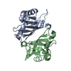
| ||||||||
|---|---|---|---|---|---|---|---|---|---|
| 1 |
| ||||||||
| Unit cell |
| ||||||||
| Details | The active form of KSI is dimer |
-Components
| #1: Protein | Mass: 14472.403 Da / Num. of mol.: 2 / Mutation: Y57S Source method: isolated from a genetically manipulated source Source: (gene. exp.) Pseudomonas putida (bacteria) / Production host: Escherichia coli (E. coli) / References: UniProt: P07445, steroid Delta-isomerase#2: Water | ChemComp-HOH / | Water Mass: 18.015 Da / Num. of mol.: 41 / Source method: isolated from a natural source / Formula: H2O Mass: 18.015 Da / Num. of mol.: 41 / Source method: isolated from a natural source / Formula: H2O |
|---|
-Experimental details
-Experiment
| Experiment | Method: X-RAY DIFFRACTION / Number of used crystals: 1 |
|---|
- Sample preparation
| Crystal | Density Matthews: 2.87 Å3/Da / Density % sol: 57.15 % | ||||||||||||||||||
|---|---|---|---|---|---|---|---|---|---|---|---|---|---|---|---|---|---|---|---|
| Crystal grow | Temperature: 295 K / Method: evaporation / pH: 4.6 Details: Sodium Acetate, Ammonium acetate, pH 4.6, EVAPORATION, temperature 295K | ||||||||||||||||||
| Crystal grow | *PLUS Temperature: 22 ℃ / Method: vapor diffusion, hanging drop | ||||||||||||||||||
| Components of the solutions | *PLUS
|
-Data collection
| Diffraction | Mean temperature: 100 K |
|---|---|
| Diffraction source | Source: ROTATING ANODE / Type: MACSCIENCE / Wavelength: 1.5418 Å |
| Detector | Type: MACSCIENCE / Detector: IMAGE PLATE / Date: Jul 18, 1997 / Details: monochromator |
| Radiation | Monochromator: Graphite / Protocol: SINGLE WAVELENGTH / Monochromatic (M) / Laue (L): M / Scattering type: x-ray |
| Radiation wavelength | Wavelength: 1.5418 Å / Relative weight: 1 |
| Reflection | Resolution: 2.2→20 Å / Num. all: 20000 / Num. obs: 15486 / % possible obs: 94.9 % / Observed criterion σ(F): 1 / Observed criterion σ(I): 1 |
| Reflection shell | Resolution: 2.2→2.4 Å / % possible all: 82 |
| Reflection | *PLUS Lowest resolution: 20 Å / Rmerge(I) obs: 0.07 |
- Processing
| Software |
| ||||||||||||||||||||
|---|---|---|---|---|---|---|---|---|---|---|---|---|---|---|---|---|---|---|---|---|---|
| Refinement | Method to determine structure: MOLECULAR REPLACEMENT / Resolution: 2.2→8 Å / σ(F): 2 / Stereochemistry target values: Engh & Huber
| ||||||||||||||||||||
| Refinement step | Cycle: LAST / Resolution: 2.2→8 Å
| ||||||||||||||||||||
| Refine LS restraints |
| ||||||||||||||||||||
| Refinement | *PLUS Lowest resolution: 8 Å / σ(F): 2 / Rfactor obs: 0.235 / Rfactor Rfree: 0.319 / Rfactor Rwork: 0.235 | ||||||||||||||||||||
| Solvent computation | *PLUS | ||||||||||||||||||||
| Displacement parameters | *PLUS |
