 Movie
Movie Controller


+ Open data

- Basic information
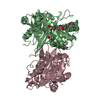
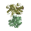
| Entry | Database: PDB / ID: 1k38 | ||||||
|---|---|---|---|---|---|---|---|
| Title | CRYSTAL STRUCTURE OF THE CLASS D BETA-LACTAMASE OXA-2 | ||||||
 Components Components | Beta-lactamase OXA-2 | ||||||
 Keywords Keywords |  HYDROLASE / serine beta-lactamase / antibiotic resistance / carbamylated lysine HYDROLASE / serine beta-lactamase / antibiotic resistance / carbamylated lysine | ||||||
| Function / homology |  Function and homology informationpenicillin binding / antibiotic catabolic process / beta-lactamase activity / beta-lactamase / response to antibiotic Function and homology informationpenicillin binding / antibiotic catabolic process / beta-lactamase activity / beta-lactamase / response to antibioticSimilarity search - Function | ||||||
| Biological species |  Salmonella typhimurium (bacteria) Salmonella typhimurium (bacteria) | ||||||
| Method | X-RAY DIFFRACTION / SYNCHROTRON / MOLECULAR REPLACEMENT / Resolution: 1.5 Å | ||||||
 Authors Authors | Kerff, F. / Fonze, E. / Bouillenne, F. / Frere, J.M. / Charlier, P. | ||||||
 Citation Citation | Journal: To be Published Title: CRYSTAL STRUCTURE OF THE CLASS D BETA-LACTAMASE OXA-2 Authors: Kerff, F. / Fonze, E. / Bouillenne, F. / Frere, J.M. / Charlier, P. #1: Journal: BIOCHEM.J. / Year: 1993Title: Substrate inactivation of the OXA2 beta-lactamase Authors: Ledent, P. / Frere, J.M. #2: Journal: BIOCHEM.J. / Year: 1993Title: A comparative study of class-D beta-lactamase Authors: Ledent, P. / Raquet, X. / Joris, B. / Van Beumen, J. / Frere, J.M. | ||||||
| History |
|
- Structure visualization
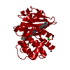
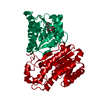
| Structure viewer | Molecule: MolmilJmol/JSmol |
|---|
- Downloads & links
-Download
| PDBx/mmCIF format | 1k38.cif.gz | 115.6 KB | Display | PDBx/mmCIF format |
|---|---|---|---|---|
| PDB format | pdb1k38.ent.gz | 89.6 KB | Display | PDB format |
| PDBx/mmJSON format | 1k38.json.gz | Tree view | PDBx/mmJSON format | |
| Others |  Other downloads Other downloads |
-Validation report
| Arichive directory | https://data.pdbj.org/pub/pdb/validation_reports/k3/1k38ftp://data.pdbj.org/pub/pdb/validation_reports/k3/1k38 | HTTPS FTP |
|---|
-Related structure data
| Related structure data |  1k4fS S: Starting model for refinement |
|---|---|
| Similar structure data |


-Links
 PDBj
PDBj- Assembly
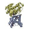
| Deposited unit | 
| ||||||||
|---|---|---|---|---|---|---|---|---|---|
| 1 |
| ||||||||
| Unit cell |
| ||||||||
| Components on special symmetry positions |
| ||||||||
| Details | the biological assembly is a monomer or a dimer depending on conditions |
-Components
| #1: Protein | Mass: 29447.264 Da / Num. of mol.: 2 Source method: isolated from a genetically manipulated source Source: (gene. exp.) Salmonella typhimurium (bacteria) / Gene: oxa-2 / Plasmid: pDML303 / Production host: Escherichia coli (E. coli) / Strain (production host): JM105 / References: UniProt: P0A1V8, beta-lactamase#2: Chemical | Formic acid  Mass: 46.025 Da / Num. of mol.: 3 / Source method: obtained synthetically / Formula: CH2O2 Mass: 46.025 Da / Num. of mol.: 3 / Source method: obtained synthetically / Formula: CH2O2#3: Water | ChemComp-HOH / | Water Mass: 18.015 Da / Num. of mol.: 314 / Source method: isolated from a natural source / Formula: H2O Mass: 18.015 Da / Num. of mol.: 314 / Source method: isolated from a natural source / Formula: H2O |
|---|
-Experimental details
-Experiment
| Experiment | Method: X-RAY DIFFRACTION / Number of used crystals: 1 |
|---|
- Sample preparation
| Crystal | Density Matthews: 2.31 Å3/Da / Density % sol: 46.84 % |
|---|---|
| Crystal grow | Temperature: 293 K / Method: vapor diffusion, hanging drop / pH: 9 Details: sodium formate, bicine, pH 9, VAPOR DIFFUSION, HANGING DROP, temperature 293K |
-Data collection
| Diffraction | Mean temperature: 100 K |
|---|---|
| Diffraction source | Source: SYNCHROTRON / Site: ESRF  / Beamline: BM14 / Wavelength: 1 Å / Beamline: BM14 / Wavelength: 1 Å |
| Detector | Type: MARRESEARCH / Detector: CCD / Date: Jun 7, 2000 |
| Radiation | Monochromator: Si 111 CHANNEL / Protocol: SINGLE WAVELENGTH / Monochromatic (M) / Laue (L): M / Scattering type: x-ray |
| Radiation wavelength | Wavelength: 1 Å / Relative weight: 1 |
| Reflection | Resolution: 1.45→25.08 Å / Num. all: 86412 / Num. obs: 86412 / % possible obs: 89.4 % / Observed criterion σ(F): 0 / Observed criterion σ(I): 0 / Redundancy: 5.4 % / Biso Wilson estimate: 19.1 Å2 / Rsym value: 0.044 / Net I/σ(I): 10.4 |
| Reflection shell | Resolution: 1.45→1.49 Å / Redundancy: 2.1 % / Mean I/σ(I) obs: 2 / Num. unique all: 3140 / Rsym value: 0.297 / % possible all: 50.7 |
- Processing
| Software |
| ||||||||||||||||||||||||||||||||||||
|---|---|---|---|---|---|---|---|---|---|---|---|---|---|---|---|---|---|---|---|---|---|---|---|---|---|---|---|---|---|---|---|---|---|---|---|---|---|
| Refinement | Method to determine structure: MOLECULAR REPLACEMENT Starting model: PDB ENTRY 1K4F, STRUCTURE OF OXA-10 Resolution: 1.5→25.08 Å / Rfactor Rfree error: 0.002 / Data cutoff high absF: 2683187.98 / Data cutoff low absF: 0 / Isotropic thermal model: RESTRAINED / Cross valid method: THROUGHOUT / σ(F): 0 / Stereochemistry target values: Engh & Huber
| ||||||||||||||||||||||||||||||||||||
| Solvent computation | Solvent model: FLAT MODEL / Bsol: 45.295 Å2 / ksol: 0.392708 e/Å3 | ||||||||||||||||||||||||||||||||||||
| Displacement parameters | Biso mean: 22.1 Å2
| ||||||||||||||||||||||||||||||||||||
| Refine analyze |
| ||||||||||||||||||||||||||||||||||||
| Refinement step | Cycle: LAST / Resolution: 1.5→25.08 Å
| ||||||||||||||||||||||||||||||||||||
| Refine LS restraints |
| ||||||||||||||||||||||||||||||||||||
| LS refinement shell | Resolution: 1.5→1.55 Å / Rfactor Rfree error: 0.013 / Total num. of bins used: 10
| ||||||||||||||||||||||||||||||||||||
| Xplor file |
|
