 Movie
Movie Controller

[English] 日本語
 Yorodumi
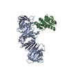
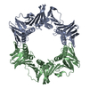
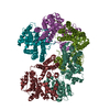
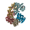
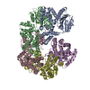
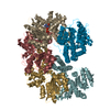
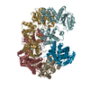
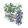
- PDB-1jr3: Crystal Structure of the Processivity Clamp Loader Gamma Complex ... -
+ Open data

- Basic information
| Entry | Database: PDB / ID: 1jr3 | ||||||
|---|---|---|---|---|---|---|---|
| Title | Crystal Structure of the Processivity Clamp Loader Gamma Complex of E. coli DNA Polymerase III | ||||||
 Components Components |
| ||||||
 Keywords Keywords |  TRANSFERASE / DNA Polymerase / Processivity / Processivity clamp / clamp loader / AAA+ ATPASE TRANSFERASE / DNA Polymerase / Processivity / Processivity clamp / clamp loader / AAA+ ATPASE | ||||||
| Function / homology |  Function and homology informationDNA polymerase III, clamp loader complex / DNA polymerase III complex / replisome / DNA polymerase processivity factor activity / 3'-5' exonuclease activity / ribonucleoside triphosphate phosphatase activity / DNA-templated DNA replication / DNA replication / DNA-directed DNA polymerase / DNA-directed DNA polymerase activity ...DNA polymerase III, clamp loader complex / DNA polymerase III complex / replisome / DNA polymerase processivity factor activity / 3'-5' exonuclease activity / ribonucleoside triphosphate phosphatase activity / DNA-templated DNA replication / DNA replication / DNA-directed DNA polymerase / DNA-directed DNA polymerase activity / ATP hydrolysis activity / DNA binding / ATP binding / identical protein binding Function and homology informationDNA polymerase III, clamp loader complex / DNA polymerase III complex / replisome / DNA polymerase processivity factor activity / 3'-5' exonuclease activity / ribonucleoside triphosphate phosphatase activity / DNA-templated DNA replication / DNA replication / DNA-directed DNA polymerase / DNA-directed DNA polymerase activity ...DNA polymerase III, clamp loader complex / DNA polymerase III complex / replisome / DNA polymerase processivity factor activity / 3'-5' exonuclease activity / ribonucleoside triphosphate phosphatase activity / DNA-templated DNA replication / DNA replication / DNA-directed DNA polymerase / DNA-directed DNA polymerase activity / ATP hydrolysis activity / DNA binding / ATP binding / identical protein bindingSimilarity search - Function | ||||||
| Biological species |  Escherichia coli (E. coli) Escherichia coli (E. coli) | ||||||
| Method | X-RAY DIFFRACTION / SYNCHROTRON / MIR / Resolution: 2.7 Å | ||||||
 Authors Authors | Jeruzalmi, D. / O'Donnell, M. / Kuriyan, J. | ||||||
 Citation Citation | Journal: Cell(Cambridge,Mass.) / Year: 2001 Title: Crystal structure of the processivity clamp loader gamma (gamma) complex of E. coli DNA polymerase III. Authors: Jeruzalmi, D. / O'Donnell, M. / Kuriyan, J. | ||||||
| History |
|
- Structure visualization
| Structure viewer | Molecule: MolmilJmol/JSmol |
|---|
- Downloads & links
-Download
| PDBx/mmCIF format | 1jr3.cif.gz | 350.6 KB | Display | PDBx/mmCIF format |
|---|---|---|---|---|
| PDB format | pdb1jr3.ent.gz | 281.9 KB | Display | PDB format |
| PDBx/mmJSON format | 1jr3.json.gz | Tree view | PDBx/mmJSON format | |
| Others |  Other downloads Other downloads |
-Validation report
| Arichive directory | https://data.pdbj.org/pub/pdb/validation_reports/jr/1jr3ftp://data.pdbj.org/pub/pdb/validation_reports/jr/1jr3 | HTTPS FTP |
|---|
-Related structure data
| Related structure data | |
|---|---|
| Similar structure data |


-Links
 PDBj
PDBj

- Assembly
| Deposited unit | 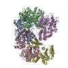
| ||||||||
|---|---|---|---|---|---|---|---|---|---|
| 1 |
| ||||||||
| Unit cell |
|
-Components
| #1: Protein | DNA polymerase III holoenzyme Mass: 41510.855 Da / Num. of mol.: 3 Source method: isolated from a genetically manipulated source Source: (gene. exp.) Escherichia coli (E. coli) / Production host: Escherichia coli (E. coli) / References: UniProt: P06710, DNA-directed DNA polymerase#2: Protein | | Mass: 38745.574 Da / Num. of mol.: 1 Source method: isolated from a genetically manipulated source Source: (gene. exp.) Escherichia coli (E. coli) / Production host: Escherichia coli (E. coli) / References: UniProt: P28630, DNA-directed DNA polymerase#3: Protein | | Mass: 36980.484 Da / Num. of mol.: 1 Source method: isolated from a genetically manipulated source Source: (gene. exp.) Escherichia coli (E. coli) / Production host: Escherichia coli (E. coli) / References: UniProt: P28631, DNA-directed DNA polymerase#4: Chemical | ChemComp-ZN /   Mass: 65.409 Da / Num. of mol.: 4 / Source method: obtained synthetically / Formula: Zn Mass: 65.409 Da / Num. of mol.: 4 / Source method: obtained synthetically / Formula: Zn#5: Chemical | Sulfate  Mass: 96.063 Da / Num. of mol.: 3 / Source method: obtained synthetically / Formula: SO4 Mass: 96.063 Da / Num. of mol.: 3 / Source method: obtained synthetically / Formula: SO4 |
|---|
-Experimental details
-Experiment
| Experiment | Method: X-RAY DIFFRACTION / Number of used crystals: 1 |
|---|
- Sample preparation
| Crystal | Density Matthews: 3.27 Å3/Da / Density % sol: 62.35 % | |||||||||||||||||||||||||||||||||||||||||||||
|---|---|---|---|---|---|---|---|---|---|---|---|---|---|---|---|---|---|---|---|---|---|---|---|---|---|---|---|---|---|---|---|---|---|---|---|---|---|---|---|---|---|---|---|---|---|---|
| Crystal grow | Temperature: 294 K / Method: vapor diffusion, hanging drop / pH: 7.5 Details: Polyethylene glycol, ammonium sulphate, dithiothreitol, dimethylacetamide, hepes, pH 7.5, VAPOR DIFFUSION, HANGING DROP, temperature 21K | |||||||||||||||||||||||||||||||||||||||||||||
| Crystal grow | *PLUS Method: unknown | |||||||||||||||||||||||||||||||||||||||||||||
| Components of the solutions | *PLUS
|
-Data collection
| Diffraction | Mean temperature: 80 K |
|---|---|
| Diffraction source | Source: SYNCHROTRON / Site: NSLS  / Beamline: X25 / Wavelength: 1.1 Å / Beamline: X25 / Wavelength: 1.1 Å |
| Detector | Type: CUSTOM-MADE / Detector: CCD / Date: Oct 17, 2000 / Details: Mirrors |
| Radiation | Protocol: SINGLE WAVELENGTH / Monochromatic (M) / Laue (L): M / Scattering type: x-ray |
| Radiation wavelength | Wavelength: 1.1 Å / Relative weight: 1 |
| Reflection | Resolution: 2.7→500 Å / Num. all: 552088 / Num. obs: 60657 / % possible obs: 83 % / Observed criterion σ(F): 2 / Observed criterion σ(I): 2 / Rsym value: 13.6 |
| Reflection shell | Resolution: 2.7→1000 Å / Num. unique all: 60657 / Rsym value: 13.6 / % possible all: 93 |
| Reflection | *PLUS Lowest resolution: 500 Å / % possible obs: 83 % / Num. measured all: 552088 / Rmerge(I) obs: 0.136 |
- Processing
| Software |
| |||||||||||||||||||||||||
|---|---|---|---|---|---|---|---|---|---|---|---|---|---|---|---|---|---|---|---|---|---|---|---|---|---|---|
| Refinement | Method to determine structure: MIR / Resolution: 2.7→500 Å / σ(F): 2 / Stereochemistry target values: Engh & Huber
| |||||||||||||||||||||||||
| Refinement step | Cycle: LAST / Resolution: 2.7→500 Å
| |||||||||||||||||||||||||
| Refine LS restraints |
| |||||||||||||||||||||||||
| Software | *PLUS Name: CNS / Version: 1 / Classification: refinement | |||||||||||||||||||||||||
| Refinement | *PLUS Highest resolution: 2.7 Å / Lowest resolution: 500 Å / σ(F): 2 / % reflection Rfree: 5 % / Rfactor obs: 0.268 | |||||||||||||||||||||||||
| Solvent computation | *PLUS | |||||||||||||||||||||||||
| Displacement parameters | *PLUS | |||||||||||||||||||||||||
| Refine LS restraints | *PLUS
|
