 Movie
Movie Controller


+ Open data

- Basic information
| Entry | Database: PDB / ID: 1jpy | |||||||||
|---|---|---|---|---|---|---|---|---|---|---|
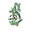
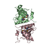
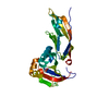
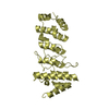
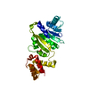
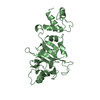
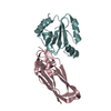
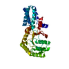
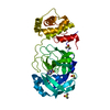
| Title | Crystal structure of IL-17F | |||||||||
 Components Components | interleukin 17F | |||||||||
 Keywords Keywords |  IMMUNE SYSTEM / cystine-knot / cytokine / t-cell derived / dimer IMMUNE SYSTEM / cystine-knot / cytokine / t-cell derived / dimer | |||||||||
| Function / homology |  Function and homology information Function and homology informationregulation of granulocyte macrophage colony-stimulating factor production / regulation of interleukin-2 production / positive regulation of lymphotoxin A production / regulation of interleukin-8 production / positive regulation of antimicrobial peptide production / Interleukin-17 signaling / positive regulation of chemokine (C-X-C motif) ligand 1 production / interleukin-17-mediated signaling pathway / regulation of transforming growth factor beta receptor signaling pathway / cytokine receptor binding ...regulation of granulocyte macrophage colony-stimulating factor production / regulation of interleukin-2 production / positive regulation of lymphotoxin A production / regulation of interleukin-8 production / positive regulation of antimicrobial peptide production / Interleukin-17 signaling / positive regulation of chemokine (C-X-C motif) ligand 1 production / interleukin-17-mediated signaling pathway / regulation of transforming growth factor beta receptor signaling pathway / cytokine receptor binding / positive regulation of cytokine production involved in inflammatory response / cartilage development / regulation of interleukin-6 production / cytokine binding / negative regulation of angiogenesis / positive regulation of cytokine production / cytokine activity / positive regulation of interleukin-6 production / Interleukin-4 and Interleukin-13 signaling / adaptive immune response / defense response to Gram-negative bacterium / defense response to Gram-positive bacterium / inflammatory response / protein heterodimerization activity / innate immune response / SARS-CoV-2 activates/modulates innate and adaptive immune responses / protein homodimerization activity / positive regulation of transcription by RNA polymerase II / extracellular space / extracellular regionSimilarity search - Function | |||||||||
| Biological species |  Homo sapiens (human) Homo sapiens (human) | |||||||||
| Method | X-RAY DIFFRACTION / SYNCHROTRON / MAD / Resolution: 2.85 Å | |||||||||
 Authors Authors | Hymowitz, S.G. / Filvaroff, E.H. / Yin, J. / Lee, J. / Cai, L. / Risser, P. / Maruoka, M. / Mao, W. / Foster, J. / Kelley, R. ...Hymowitz, S.G. / Filvaroff, E.H. / Yin, J. / Lee, J. / Cai, L. / Risser, P. / Maruoka, M. / Mao, W. / Foster, J. / Kelley, R. / Pan, G. / Gurney, A.L. / de Vos, A.M. / Starovasnik, M.A. | |||||||||
 Citation Citation | Journal: EMBO J. / Year: 2001 Title: IL-17s adopt a cystine knot fold: structure and activity of a novel cytokine, IL-17F, and implications for receptor binding. Authors: Hymowitz, S.G. / Filvaroff, E.H. / Yin, J.P. / Lee, J. / Cai, L. / Risser, P. / Maruoka, M. / Mao, W. / Foster, J. / Kelley, R.F. / Pan, G. / Gurney, A.L. / de Vos, A.M. / Starovasnik, M.A. | |||||||||
| History |
|
- Structure visualization
| Structure viewer | Molecule: MolmilJmol/JSmol |
|---|
- Downloads & links
-Download
| PDBx/mmCIF format | 1jpy.cif.gz | 102.9 KB | Display | PDBx/mmCIF format |
|---|---|---|---|---|
| PDB format | pdb1jpy.ent.gz | 83.8 KB | Display | PDB format |
| PDBx/mmJSON format | 1jpy.json.gz | Tree view | PDBx/mmJSON format | |
| Others |  Other downloads Other downloads |
-Validation report
| Arichive directory | https://data.pdbj.org/pub/pdb/validation_reports/jp/1jpyftp://data.pdbj.org/pub/pdb/validation_reports/jp/1jpy | HTTPS FTP |
|---|
-Related structure data
| Similar structure data |
|---|

-Links
 PDBj
PDBj

- Assembly
| Deposited unit | 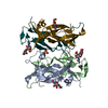
| ||||||||||
|---|---|---|---|---|---|---|---|---|---|---|---|
| 1 | 
| ||||||||||
| 2 | 
| ||||||||||
| Unit cell |
| ||||||||||
| Details | The biologically active unit is a dimer. The assymetric unit is formed of two dimers. |
-Components
-Protein , 1 types, 4 molecules ABXY
| #1: Protein | Mass: 15335.575 Da / Num. of mol.: 4 / Fragment: Secreted IL-17F Source method: isolated from a genetically manipulated source Details: the first four residues, GSHM, are from the vector / Source: (gene. exp.) Homo sapiens (human) / Gene: IL-17FPlasmid details: A modified plasmid was used which incorporated an N-terminal his tag and thrombin site under the control of the viral coat protein promotor Production host:  unidentified baculovirus / Strain (production host): Hi5 cells / References: GenBank: 15077800, UniProt: Q96PD4*PLUS unidentified baculovirus / Strain (production host): Hi5 cells / References: GenBank: 15077800, UniProt: Q96PD4*PLUS |
|---|
-Sugars , 3 types, 4 molecules 
| #2: Polysaccharide | 2-acetamido-2-deoxy-alpha-D-glucopyranose-(1-4)-2-acetamido-2-deoxy-beta-D-glucopyranose / Mass: 424.401 Da / Num. of mol.: 1 Source method: isolated from a genetically manipulated source |
|---|---|
| #3: Polysaccharide | 2-acetamido-2-deoxy-beta-D-glucopyranose-(1-4)-2-acetamido-2-deoxy-beta-D-glucopyranose / Mass: 424.401 Da / Num. of mol.: 1 Source method: isolated from a genetically manipulated source |
| #4: Sugar | N-Acetylglucosamine Type: D-saccharide, beta linking / Mass: 221.208 Da / Num. of mol.: 2 Type: D-saccharide, beta linking / Mass: 221.208 Da / Num. of mol.: 2Source method: isolated from a genetically manipulated source Formula: C8H15NO6 |
-Non-polymers , 2 types, 28 molecules 

| #5: Chemical | ChemComp-SO4 / Sulfate Mass: 96.063 Da / Num. of mol.: 1 / Source method: obtained synthetically / Formula: SO4 Mass: 96.063 Da / Num. of mol.: 1 / Source method: obtained synthetically / Formula: SO4 |
|---|---|
| #6: Water | ChemComp-HOH / WaterMass: 18.015 Da / Num. of mol.: 27 / Source method: isolated from a natural source / Formula: H2O |
-Experimental details
-Experiment
| Experiment | Method: X-RAY DIFFRACTION / Number of used crystals: 1 |
|---|
- Sample preparation
| Crystal | Density Matthews: 3.3 Å3/Da / Density % sol: 64 % | |||||||||||||||||||||||||
|---|---|---|---|---|---|---|---|---|---|---|---|---|---|---|---|---|---|---|---|---|---|---|---|---|---|---|
| Crystal grow | Temperature: 292 K / Method: vapor diffusion, hanging drop / pH: 5.6 Details: well solution consisted of 1.0M Lithium Sulfate, 0.5M Ammonium Sulfate, 100mM Sodium Citrate, 1% Ethanol at 5.6, VAPOR DIFFUSION, HANGING DROP at 292K | |||||||||||||||||||||||||
| Crystal grow | *PLUS Temperature: 19 ℃ | |||||||||||||||||||||||||
| Components of the solutions | *PLUS
|
-Data collection
| Diffraction | Mean temperature: 100 K |
|---|---|
| Diffraction source | Source: SYNCHROTRON / Site: SSRL  / Beamline: BL9-2 / Wavelength: 0.979 Å / Beamline: BL9-2 / Wavelength: 0.979 Å |
| Detector | Type: ADSC QUANTUM 4 / Detector: CCD / Date: Mar 13, 2001 |
| Radiation | Monochromator: SSRL beam line 9-2 / Protocol: MAD / Monochromatic (M) / Laue (L): M / Scattering type: x-ray |
| Radiation wavelength | Wavelength: 0.979 Å / Relative weight: 1 |
| Reflection | Resolution: 2.85→50 Å / Num. all: 19294 / Num. obs: 19294 / % possible obs: 100 % / Observed criterion σ(F): 0 / Observed criterion σ(I): -3 / Redundancy: 7.3 % / Biso Wilson estimate: 63.8 Å2 / Rmerge(I) obs: 0.088 / Rsym value: 0.088 / Net I/σ(I): 7.7 |
| Reflection shell | Resolution: 2.85→2.95 Å / Redundancy: 7.3 % / Rmerge(I) obs: 0.541 / Num. unique all: 1912 / Rsym value: 0.541 / % possible all: 100 |
| Reflection | *PLUS % possible obs: 100 % / Num. measured all: 141778 |
| Reflection shell | *PLUS % possible obs: 100 % |
- Processing
| Software |
| ||||||||||||||||||||||||||||||||||||||||
|---|---|---|---|---|---|---|---|---|---|---|---|---|---|---|---|---|---|---|---|---|---|---|---|---|---|---|---|---|---|---|---|---|---|---|---|---|---|---|---|---|---|
| Refinement | Method to determine structure: MAD / Resolution: 2.85→30 Å / Rfactor Rfree error: 0.008 / Data cutoff high absF: 10000000 / Data cutoff low absF: 0 / Isotropic thermal model: RESTRAINED / Cross valid method: THROUGHOUT / σ(F): 0.2 / σ(I): 0 / Stereochemistry target values: Engh & Huber Details: The structure was solved using data collected at three different wavelenghts from crystals that had been soaked in the Hg containing compound thimerosal but was refined against a native data ...Details: The structure was solved using data collected at three different wavelenghts from crystals that had been soaked in the Hg containing compound thimerosal but was refined against a native data set collected on a crystal that was not derivatized. The crystollgraphic statistics presented here are for the native data set. Used maximum likelihood residual. The program REFMAC was used at the initial stages of refinement. A bulk solvent corrections was applied in Xplor98.1 as part of the refinement process. This correction has not been applied to the depositted structure factors
| ||||||||||||||||||||||||||||||||||||||||
| Solvent computation | Solvent model: BULK SOLVENT MODEL | ||||||||||||||||||||||||||||||||||||||||
| Displacement parameters | Biso mean: 47.9 Å2
| ||||||||||||||||||||||||||||||||||||||||
| Refine analyze |
| ||||||||||||||||||||||||||||||||||||||||
| Refinement step | Cycle: LAST / Resolution: 2.85→30 Å
| ||||||||||||||||||||||||||||||||||||||||
| Refine LS restraints |
| ||||||||||||||||||||||||||||||||||||||||
| LS refinement shell | Resolution: 2.85→3.03 Å / Rfactor Rfree error: 0.028 / Total num. of bins used: 6
| ||||||||||||||||||||||||||||||||||||||||
| Xplor file |
| ||||||||||||||||||||||||||||||||||||||||
| Software | *PLUS Name: X-PLOR / Version: 98.1 / Classification: refinement | ||||||||||||||||||||||||||||||||||||||||
| Refinement | *PLUS σ(F): 2 / % reflection Rfree: 5.9 % | ||||||||||||||||||||||||||||||||||||||||
| Solvent computation | *PLUS | ||||||||||||||||||||||||||||||||||||||||
| Displacement parameters | *PLUS Biso mean: 47.9 Å2 | ||||||||||||||||||||||||||||||||||||||||
| Refine LS restraints | *PLUS
| ||||||||||||||||||||||||||||||||||||||||
| LS refinement shell | *PLUS Rfactor Rfree: 0.377 / % reflection Rfree: 5.6 % / Rfactor Rwork: 0.318 |
