 Movie
Movie Controller

[English] 日本語
 Yorodumi
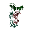
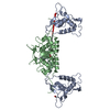
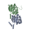
- PDB-1jps: Crystal structure of tissue factor in complex with humanized Fab D3h44 -
+ Open data

- Basic information
| Entry | Database: PDB / ID: 1jps | ||||||
|---|---|---|---|---|---|---|---|
| Title | Crystal structure of tissue factor in complex with humanized Fab D3h44 | ||||||
 Components Components |
| ||||||
 Keywords Keywords |  IMMUNE SYSTEM / antigen-antibody recognition / humanized antibody / blood coagulation / interface water molecules IMMUNE SYSTEM / antigen-antibody recognition / humanized antibody / blood coagulation / interface water molecules | ||||||
| Function / homology |  Function and homology information Function and homology informationactivation of plasma proteins involved in acute inflammatory response / activation of blood coagulation via clotting cascade / serine-type peptidase complex / positive regulation of platelet-derived growth factor receptor signaling pathway / NGF-stimulated transcription / positive regulation of positive chemotaxis / Extrinsic Pathway of Fibrin Clot Formation / cytokine receptor activity / positive regulation of TOR signaling / positive regulation of endothelial cell proliferation ...activation of plasma proteins involved in acute inflammatory response / activation of blood coagulation via clotting cascade / serine-type peptidase complex / positive regulation of platelet-derived growth factor receptor signaling pathway / NGF-stimulated transcription / positive regulation of positive chemotaxis / Extrinsic Pathway of Fibrin Clot Formation / cytokine receptor activity / positive regulation of TOR signaling / positive regulation of endothelial cell proliferation / positive regulation of interleukin-8 production / phospholipid binding / protein processing / cytokine-mediated signaling pathway / positive regulation of angiogenesis / activation of cysteine-type endopeptidase activity involved in apoptotic process / blood coagulation / collagen-containing extracellular matrix / protease binding / positive regulation of cell migration / external side of plasma membrane / positive regulation of gene expression / cell surface / extracellular space / membrane / plasma membraneSimilarity search - Function | ||||||
| Biological species |  Homo sapiens (human) Homo sapiens (human) | ||||||
| Method | X-RAY DIFFRACTION / SYNCHROTRON / MOLECULAR REPLACEMENT / Resolution: 1.85 Å | ||||||
 Authors Authors | Faelber, K. / Kirchhofer, D. / Presta, L. / Kelley, R.F. / Muller, Y.A. | ||||||
 Citation Citation | Journal: J.Mol.Biol. / Year: 2001 Title: The 1.85 A resolution crystal structures of tissue factor in complex with humanized Fab D3h44 and of free humanized Fab D3h44: revisiting the solvation of antigen combining sites. Authors: Faelber, K. / Kirchhofer, D. / Presta, L. / Kelley, R.F. / Muller, Y.A. #1: Journal: THROMB.HAEMOST. / Year: 2001Title: Generation of a humanized, high affinity anti-tissue factor antibody for use as a novel antithrombotic therapeutic Authors: Presta, L. / Sims, P. / Meng, Y.G. / Moran, P. / Bullens, S. / Bunting, S. / Schoenfeld, J. / Lowe, D. / Lai, J. / Rancatore, P. / Iverson, M. / Lim, A. / Chisholm, V. / Kelley, R.F. / ...Authors: Presta, L. / Sims, P. / Meng, Y.G. / Moran, P. / Bullens, S. / Bunting, S. / Schoenfeld, J. / Lowe, D. / Lai, J. / Rancatore, P. / Iverson, M. / Lim, A. / Chisholm, V. / Kelley, R.F. / Riederer, M. / Kirchhofer, D. #2: Journal: J.Mol.Biol. / Year: 1996Title: The crystal structure of the extracellulare domain of human tissue factor refined to 1.7 A resolution Authors: Muller, Y.A. / Ultsch, M.H. / de Vos, A.M. | ||||||
| History |
|
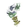
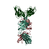
- Structure visualization
| Structure viewer | Molecule: MolmilJmol/JSmol |
|---|
- Downloads & links
-Download
| PDBx/mmCIF format | 1jps.cif.gz | 147.4 KB | Display | PDBx/mmCIF format |
|---|---|---|---|---|
| PDB format | pdb1jps.ent.gz | 113.1 KB | Display | PDB format |
| PDBx/mmJSON format | 1jps.json.gz | Tree view | PDBx/mmJSON format | |
| Others |  Other downloads Other downloads |
-Validation report
| Arichive directory | https://data.pdbj.org/pub/pdb/validation_reports/jp/1jpsftp://data.pdbj.org/pub/pdb/validation_reports/jp/1jps | HTTPS FTP |
|---|
-Related structure data
| Related structure data | 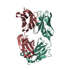 1jptC 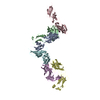 1ahwS C: citing same article ( S: Starting model for refinement |
|---|---|
| Similar structure data |


-Links
 PDBj
PDBj


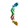
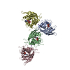
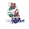
- Assembly
| Deposited unit | 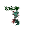
| ||||||||
|---|---|---|---|---|---|---|---|---|---|
| 1 |
| ||||||||
| Unit cell |
|
-Components
| #1: Antibody | Mass: 23523.107 Da / Num. of mol.: 1 / Fragment: Fab fragment Source method: isolated from a genetically manipulated source Source: (gene. exp.) Homo sapiens (human) / Plasmid: pEMX1 / Production host:  Escherichia coli (E. coli) Escherichia coli (E. coli) |
|---|---|
| #2: Antibody | Mass: 24043.805 Da / Num. of mol.: 1 / Fragment: Fab fragment Source method: isolated from a genetically manipulated source Source: (gene. exp.) Homo sapiens (human) / Plasmid: pEMX1 / Production host: Escherichia coli (E. coli) |
| #3: Protein | Mass: 24826.512 Da / Num. of mol.: 1 / Fragment: extracellular domain Source method: isolated from a genetically manipulated source Source: (gene. exp.) Homo sapiens (human) / Plasmid: pEMX1 / Production host: Escherichia coli (E. coli) / References: UniProt: P13726 |
| #4: Water | ChemComp-HOH / Water Mass: 18.015 Da / Num. of mol.: 579 / Source method: isolated from a natural source / Formula: H2O Mass: 18.015 Da / Num. of mol.: 579 / Source method: isolated from a natural source / Formula: H2O |
-Experimental details
-Experiment
| Experiment | Method: X-RAY DIFFRACTION / Number of used crystals: 1 |
|---|
- Sample preparation
| Crystal | Density Matthews: 2.98 Å3/Da / Density % sol: 58.78 % | |||||||||||||||||||||||||||||||||||||||||||||||||
|---|---|---|---|---|---|---|---|---|---|---|---|---|---|---|---|---|---|---|---|---|---|---|---|---|---|---|---|---|---|---|---|---|---|---|---|---|---|---|---|---|---|---|---|---|---|---|---|---|---|---|
| Crystal grow | Temperature: 277 K / Method: vapor diffusion, hanging drop / pH: 7.5 Details: PEG 6000, sodium chloride, sodium-HEPES buffer, pH 7.5, VAPOR DIFFUSION, HANGING DROP, temperature 277K | |||||||||||||||||||||||||||||||||||||||||||||||||
| Crystal grow | *PLUS Temperature: 4 ℃ / Method: vapor diffusion | |||||||||||||||||||||||||||||||||||||||||||||||||
| Components of the solutions | *PLUS
|
-Data collection
| Diffraction | Mean temperature: 110 K |
|---|---|
| Diffraction source | Source: SYNCHROTRON / Site: EMBL/DESY, HAMBURG  / Beamline: BW7B / Wavelength: 0.8428 Å / Beamline: BW7B / Wavelength: 0.8428 Å |
| Detector | Type: MARRESEARCH / Detector: IMAGE PLATE / Date: May 18, 2000 / Details: mirrors |
| Radiation | Monochromator: triangular monochromator / Protocol: SINGLE WAVELENGTH / Monochromatic (M) / Laue (L): M / Scattering type: x-ray |
| Radiation wavelength | Wavelength: 0.8428 Å / Relative weight: 1 |
| Reflection | Resolution: 1.85→40 Å / Num. all: 73056 / Num. obs: 73056 / % possible obs: 96.8 % / Observed criterion σ(F): 0 / Observed criterion σ(I): 0 / Redundancy: 3.4 % / Biso Wilson estimate: 30.1 Å2 / Rsym value: 0.061 / Net I/σ(I): 12.6 |
| Reflection shell | Resolution: 1.85→1.96 Å / Redundancy: 3.5 % / Mean I/σ(I) obs: 3.7 / Num. unique all: 11059 / Rsym value: 0.27 / % possible all: 94.4 |
- Processing
| Software |
| |||||||||||||||||||||||||
|---|---|---|---|---|---|---|---|---|---|---|---|---|---|---|---|---|---|---|---|---|---|---|---|---|---|---|
| Refinement | Method to determine structure: MOLECULAR REPLACEMENT Starting model: PDB ENTRY 1AHW Resolution: 1.85→40 Å / Cross valid method: THROUGHOUT / σ(F): 0 / σ(I): 0 / Stereochemistry target values: Engh & Huber
| |||||||||||||||||||||||||
| Displacement parameters | Biso mean: 29.2 Å2
| |||||||||||||||||||||||||
| Refinement step | Cycle: LAST / Resolution: 1.85→40 Å
| |||||||||||||||||||||||||
| Refine LS restraints |
| |||||||||||||||||||||||||
| LS refinement shell | Resolution: 1.85→1.97 Å / Rfactor Rfree error: 0.01
| |||||||||||||||||||||||||
| Software | *PLUS Name: CNS / Version: 1 / Classification: refinement | |||||||||||||||||||||||||
| Refinement | *PLUS Lowest resolution: 40 Å / Num. reflection obs: 67779 / σ(F): 0 / % reflection Rfree: 7.221 % / Rfactor obs: 0.201 / Rfactor Rfree: 0.225 | |||||||||||||||||||||||||
| Solvent computation | *PLUS | |||||||||||||||||||||||||
| Displacement parameters | *PLUS Biso mean: 29.2 Å2 | |||||||||||||||||||||||||
| LS refinement shell | *PLUS Rfactor Rfree: 0.278 / Rfactor Rwork: 0.256 |
