 Movie
Movie Controller

[English] 日本語
 Yorodumi
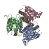
- PDB-1jlj: 1.6 Angstrom crystal structure of the human neuroreceptor anchori... -
+ Open data

- Basic information
| Entry | Database: PDB / ID: 1jlj | ||||||
|---|---|---|---|---|---|---|---|
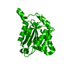
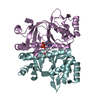
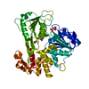
| Title | 1.6 Angstrom crystal structure of the human neuroreceptor anchoring and molybdenum cofactor biosynthesis protein gephyrin | ||||||
 Components Components | gephyrin | ||||||
 Keywords Keywords | STRUCTURAL PROTEIN / globular alpha/beta fold | ||||||
| Function / homology |  Function and homology information Function and homology informationmolybdenum incorporation into molybdenum-molybdopterin complex / : / Molybdenum cofactor biosynthesis / glycine receptor clustering / molybdopterin cofactor biosynthetic process / establishment of synaptic specificity at neuromuscular junction / molybdopterin adenylyltransferase / molybdopterin adenylyltransferase activity / gamma-aminobutyric acid receptor clustering / molybdopterin molybdotransferase ...molybdenum incorporation into molybdenum-molybdopterin complex / : / Molybdenum cofactor biosynthesis / glycine receptor clustering / molybdopterin cofactor biosynthetic process / establishment of synaptic specificity at neuromuscular junction / molybdopterin adenylyltransferase / molybdopterin adenylyltransferase activity / gamma-aminobutyric acid receptor clustering / molybdopterin molybdotransferase / molybdopterin molybdotransferase activity / postsynaptic specialization / nitrate reductase activity / Mo-molybdopterin cofactor biosynthetic process / postsynaptic neurotransmitter receptor diffusion trapping / postsynaptic specialization membrane / response to metal ion / molybdopterin cofactor binding / postsynaptic membrane / postsynaptic density / cytoskeleton / dendrite / ATP binding / metal ion binding / plasma membrane / cytosol / cytoplasmSimilarity search - Function | ||||||
| Biological species |  Homo sapiens (human) Homo sapiens (human) | ||||||
| Method | X-RAY DIFFRACTION / SYNCHROTRON / MOLECULAR REPLACEMENT / Resolution: 1.6 Å | ||||||
 Authors Authors | Schwarz, G. / Schrader, N. / Mendel, R.R. / Hecht, H.-J. / Schindelin, H. | ||||||
 Citation Citation | Journal: J.Mol.Biol. / Year: 2001 Title: Crystal structures of human gephyrin and plant Cnx1 G domains: comparative analysis and functional implications. Authors: Schwarz, G. / Schrader, N. / Mendel, R.R. / Hecht, H.J. / Schindelin, H. | ||||||
| History |
|
- Structure visualization
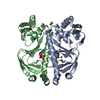
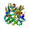
| Structure viewer | Molecule: MolmilJmol/JSmol |
|---|
- Downloads & links
-Download
| PDBx/mmCIF format | 1jlj.cif.gz | 128.8 KB | Display | PDBx/mmCIF format |
|---|---|---|---|---|
| PDB format | pdb1jlj.ent.gz | 98.7 KB | Display | PDB format |
| PDBx/mmJSON format | 1jlj.json.gz | Tree view | PDBx/mmJSON format | |
| Others |  Other downloads Other downloads |
-Validation report
| Arichive directory | https://data.pdbj.org/pub/pdb/validation_reports/jl/1jljftp://data.pdbj.org/pub/pdb/validation_reports/jl/1jlj | HTTPS FTP |
|---|
-Related structure data
| Related structure data |  1eavC  1di6S S: Starting model for refinement C: citing same article ( |
|---|---|
| Similar structure data |


-Links
 PDBj
PDBj- Assembly
| Deposited unit | 
| ||||||||||
|---|---|---|---|---|---|---|---|---|---|---|---|
| 1 |
| ||||||||||
| Unit cell |
| ||||||||||
| Components on special symmetry positions |
| ||||||||||
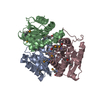
| Details | content of the assymetric unit corresponds to the biological active form of the trimeric gephyrin G domain |
-Components
| #1: Protein | / PUTATIVE GLYCINE RECEPTOR-TUBULIN LINKER PROTEIN Mass: 20812.020 Da / Num. of mol.: 3 / Fragment: residues 1-181 Source method: isolated from a genetically manipulated source Source: (gene. exp.) Homo sapiens (human) / Gene: GEPHYRIN / Plasmid: pQE60 / Production host:  Escherichia coli (E. coli) / Strain (production host): DL41 / References: UniProt: Q9NQX3 Escherichia coli (E. coli) / Strain (production host): DL41 / References: UniProt: Q9NQX3#2: Chemical | ChemComp-NA / |   Mass: 22.990 Da / Num. of mol.: 1 / Source method: obtained synthetically / Formula: Na Mass: 22.990 Da / Num. of mol.: 1 / Source method: obtained synthetically / Formula: Na#3: Chemical | ChemComp-FMT / | Formic acid  Mass: 46.025 Da / Num. of mol.: 1 / Source method: obtained synthetically / Formula: CH2O2 Mass: 46.025 Da / Num. of mol.: 1 / Source method: obtained synthetically / Formula: CH2O2#4: Water | ChemComp-HOH / | Water Mass: 18.015 Da / Num. of mol.: 770 / Source method: isolated from a natural source / Formula: H2O Mass: 18.015 Da / Num. of mol.: 770 / Source method: isolated from a natural source / Formula: H2O |
|---|
-Experimental details
-Experiment
| Experiment | Method: X-RAY DIFFRACTION / Number of used crystals: 2 |
|---|
- Sample preparation
| Crystal | Density Matthews: 2.2 Å3/Da / Density % sol: 44.19 % | ||||||||||||||||||||
|---|---|---|---|---|---|---|---|---|---|---|---|---|---|---|---|---|---|---|---|---|---|
| Crystal grow | Temperature: 294 K / Method: vapor diffusion, hanging drop / pH: 4.6 Details: sodium formate, ammonium, acetate, pH 4.6, VAPOR DIFFUSION, HANGING DROP at 294K | ||||||||||||||||||||
| Crystal grow | *PLUS Method: vapor diffusion | ||||||||||||||||||||
| Components of the solutions | *PLUS
|
-Data collection
| Diffraction |
| ||||||||||||||||||
|---|---|---|---|---|---|---|---|---|---|---|---|---|---|---|---|---|---|---|---|
| Diffraction source |
| ||||||||||||||||||
| Detector |
| ||||||||||||||||||
| Radiation |
| ||||||||||||||||||
| Radiation wavelength | Wavelength: 1.1 Å / Relative weight: 1 | ||||||||||||||||||
| Reflection | Resolution: 1.6→50 Å / Num. all: 71681 / Num. obs: 64743 / % possible obs: 90.9 % / Observed criterion σ(F): 0 / Observed criterion σ(I): 0 / Redundancy: 3.7 % / Rsym value: 0.116 / Net I/σ(I): 14.4 | ||||||||||||||||||
| Reflection shell | Resolution: 1.6→1.66 Å / Mean I/σ(I) obs: 1.9 / Rsym value: 0.283 / % possible all: 46.3 | ||||||||||||||||||
| Reflection | *PLUS Lowest resolution: 50 Å / Rmerge(I) obs: 0.116 | ||||||||||||||||||
| Reflection shell | *PLUS % possible obs: 46.3 % / Rmerge(I) obs: 0.283 |

- Processing
| Software |
| ||||||||||||||||||||||||||||
|---|---|---|---|---|---|---|---|---|---|---|---|---|---|---|---|---|---|---|---|---|---|---|---|---|---|---|---|---|---|
| Refinement | Method to determine structure: MOLECULAR REPLACEMENT Starting model: PDB ENTRY 1DI6 Resolution: 1.6→20 Å / Isotropic thermal model: Isotropic / Cross valid method: THROUGHOUT / σ(F): 0 / Stereochemistry target values: REFMAC library Details: Scaling details: Babinet's principle for scaling has been used. Bulk solvent correction based on constant value has been used. Parameters for mask calculation. VDW prob radii = 1.40, ION ...Details: Scaling details: Babinet's principle for scaling has been used. Bulk solvent correction based on constant value has been used. Parameters for mask calculation. VDW prob radii = 1.40, ION probe radii = 0.80, Shrinkage radii = 0.80
| ||||||||||||||||||||||||||||
| Solvent computation | Solvent model: bulk solvent correction | ||||||||||||||||||||||||||||
| Displacement parameters | Biso mean: 18.828 Å2
| ||||||||||||||||||||||||||||
| Refinement step | Cycle: LAST / Resolution: 1.6→20 Å
| ||||||||||||||||||||||||||||
| Refine LS restraints |
| ||||||||||||||||||||||||||||
| Software | *PLUS Name: REFMAC / Classification: refinement | ||||||||||||||||||||||||||||
| Refinement | *PLUS Highest resolution: 1.6 Å / Lowest resolution: 20 Å / σ(F): 0 / % reflection Rfree: 4.1 % | ||||||||||||||||||||||||||||
| Solvent computation | *PLUS | ||||||||||||||||||||||||||||
| Displacement parameters | *PLUS | ||||||||||||||||||||||||||||
| Refine LS restraints | *PLUS
|
