 Movie
Movie Controller


+ Open data

- Basic information
| Entry | Database: PDB / ID: 1jlh | ||||||
|---|---|---|---|---|---|---|---|
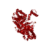
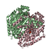
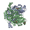
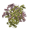
| Title | Human Glucose-6-phosphate Isomerase | ||||||
 Components Components | phosphoglucose isomerase Glucose-6-phosphate isomerase Glucose-6-phosphate isomerase | ||||||
 Keywords Keywords | ISOMERASE / glycolysis / glyconeogenesis | ||||||
| Function / homology |  Function and homology informationglucose-6-phosphate isomerase / glucose-6-phosphate isomerase activity / hemostasis / glucose 6-phosphate metabolic process / carbohydrate derivative binding / Gluconeogenesis / monosaccharide binding / Glycolysis / erythrocyte homeostasis / ciliary membrane ...glucose-6-phosphate isomerase / glucose-6-phosphate isomerase activity / hemostasis / glucose 6-phosphate metabolic process / carbohydrate derivative binding / Gluconeogenesis / monosaccharide binding / Glycolysis / erythrocyte homeostasis / ciliary membrane / positive regulation of immunoglobulin production / response to testosterone / humoral immune response / mesoderm formation / response to immobilization stress / response to cadmium ion / response to muscle stretch / positive regulation of endothelial cell migration / response to progesterone / cytokine activity / gluconeogenesis / TP53 Regulates Metabolic Genes / glycolytic process / growth factor activity / response to estradiol / glucose homeostasis / secretory granule lumen / in utero embryonic development / ficolin-1-rich granule lumen / negative regulation of neuron apoptotic process / learning or memory / carbohydrate metabolic process / ubiquitin protein ligase binding / Neutrophil degranulation / extracellular exosome / extracellular region / membrane / cytosol Function and homology informationglucose-6-phosphate isomerase / glucose-6-phosphate isomerase activity / hemostasis / glucose 6-phosphate metabolic process / carbohydrate derivative binding / Gluconeogenesis / monosaccharide binding / Glycolysis / erythrocyte homeostasis / ciliary membrane ...glucose-6-phosphate isomerase / glucose-6-phosphate isomerase activity / hemostasis / glucose 6-phosphate metabolic process / carbohydrate derivative binding / Gluconeogenesis / monosaccharide binding / Glycolysis / erythrocyte homeostasis / ciliary membrane / positive regulation of immunoglobulin production / response to testosterone / humoral immune response / mesoderm formation / response to immobilization stress / response to cadmium ion / response to muscle stretch / positive regulation of endothelial cell migration / response to progesterone / cytokine activity / gluconeogenesis / TP53 Regulates Metabolic Genes / glycolytic process / growth factor activity / response to estradiol / glucose homeostasis / secretory granule lumen / in utero embryonic development / ficolin-1-rich granule lumen / negative regulation of neuron apoptotic process / learning or memory / carbohydrate metabolic process / ubiquitin protein ligase binding / Neutrophil degranulation / extracellular exosome / extracellular region / membrane / cytosolSimilarity search - Function | ||||||
| Biological species |  Homo sapiens (human) Homo sapiens (human) | ||||||
| Method | X-RAY DIFFRACTION / SYNCHROTRON / MOLECULAR REPLACEMENT / Resolution: 2.1 Å | ||||||
 Authors Authors | Cordeiro, A.T. | ||||||
 Citation Citation | Journal: BIOCHIM.BIOPHYS.ACTA / Year: 2003 Title: Crystal structure of human phosphoglucose isomerase and analysis of the initial catalytic steps Authors: Cordeiro, A.T. / Godoi, P.H.C. / Silva, C.H.T.P. / Garratt, R.C. / Oliva, G. / Thiemann, O.H. | ||||||
| History |
|
- Structure visualization
| Structure viewer | Molecule: MolmilJmol/JSmol |
|---|
- Downloads & links
-Download
| PDBx/mmCIF format | 1jlh.cif.gz | 462 KB | Display | PDBx/mmCIF format |
|---|---|---|---|---|
| PDB format | pdb1jlh.ent.gz | 379 KB | Display | PDB format |
| PDBx/mmJSON format | 1jlh.json.gz | Tree view | PDBx/mmJSON format | |
| Others |  Other downloads Other downloads |
-Validation report
| Arichive directory | https://data.pdbj.org/pub/pdb/validation_reports/jl/1jlhftp://data.pdbj.org/pub/pdb/validation_reports/jl/1jlh | HTTPS FTP |
|---|
-Related structure data
| Related structure data |  1dqrS S: Starting model for refinement |
|---|---|
| Similar structure data |


-Links
 PDBj
PDBj


- Assembly
| Deposited unit | 
| ||||||||
|---|---|---|---|---|---|---|---|---|---|
| 1 | 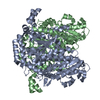
| ||||||||
| 2 | 
| ||||||||
| Unit cell |
|
-Components
| #1: Protein | Glucose-6-phosphate isomerase / PGI / GLUCOSE PHOSPHATE ISOMERASE Mass: 63229.879 Da / Num. of mol.: 4 Source method: isolated from a genetically manipulated source Source: (gene. exp.) Homo sapiens (human) / Plasmid: pET29-a(+) / Species (production host): Escherichia coli / Production host:  Escherichia coli BL21(DE3) (bacteria) / Strain (production host): BL21(DE3) / References: UniProt: P06744, glucose-6-phosphate isomerase Escherichia coli BL21(DE3) (bacteria) / Strain (production host): BL21(DE3) / References: UniProt: P06744, glucose-6-phosphate isomerase#2: Water | ChemComp-HOH / | Water Mass: 18.015 Da / Num. of mol.: 1289 / Source method: isolated from a natural source / Formula: H2O Mass: 18.015 Da / Num. of mol.: 1289 / Source method: isolated from a natural source / Formula: H2O |
|---|
-Experimental details
-Experiment
| Experiment | Method: X-RAY DIFFRACTION / Number of used crystals: 1 |
|---|
- Sample preparation
| Crystal | Density Matthews: 2.41 Å3/Da / Density % sol: 48.64 % | ||||||||||||||||||||||||
|---|---|---|---|---|---|---|---|---|---|---|---|---|---|---|---|---|---|---|---|---|---|---|---|---|---|
| Crystal grow | Temperature: 291 K / Method: vapor diffusion, hanging drop / pH: 7.5 Details: 10% PEG 10000, 0.1M HEPES , pH 7.5, VAPOR DIFFUSION, HANGING DROP, temperature 291K | ||||||||||||||||||||||||
| Crystal grow | *PLUS Details: Cordeiro, A.T., (2001) Acta Crystallogr., D57, 592. | ||||||||||||||||||||||||
| Components of the solutions | *PLUS
|
-Data collection
| Diffraction | Mean temperature: 100 K |
|---|---|
| Diffraction source | Source: SYNCHROTRON / Site: LNLS  / Beamline: D03B-MX1 / Wavelength: 1.545 Å / Beamline: D03B-MX1 / Wavelength: 1.545 Å |
| Detector | Type: MAR scanner 345 mm plate / Detector: IMAGE PLATE / Date: Jul 1, 2000 |
| Radiation | Protocol: SINGLE WAVELENGTH / Monochromatic (M) / Laue (L): M / Scattering type: x-ray |
| Radiation wavelength | Wavelength: 1.545 Å / Relative weight: 1 |
| Reflection | Resolution: 2.1→22.21 Å / Num. all: 138847 / Num. obs: 138362 / % possible obs: 99.6 % / Observed criterion σ(F): 0 / Observed criterion σ(I): 0 / Redundancy: 4.8 % / Biso Wilson estimate: 15.5 Å2 / Limit h max: 38 / Limit h min: 0 / Limit k max: 51 / Limit k min: 0 / Limit l max: 129 / Limit l min: 0 / Observed criterion F max: 3704005.62 / Observed criterion F min: 24.9 / Rmerge(I) obs: 0.111 / Net I/σ(I): 6.7 |
| Reflection shell | Resolution: 2.1→2.23 Å / Rmerge(I) obs: 0.399 / % possible all: 99.4 |
| Reflection | *PLUS Highest resolution: 2.1 Å / % possible obs: 99.7 % / Num. measured all: 672976 |
| Reflection shell | *PLUS Highest resolution: 2.1 Å / % possible obs: 99.4 % |
- Processing
| Software |
| |||||||||||||||||||||||||||||||||||||||||||||||||||||||||||||||||||||||||||||||||||||||||||||||||||
|---|---|---|---|---|---|---|---|---|---|---|---|---|---|---|---|---|---|---|---|---|---|---|---|---|---|---|---|---|---|---|---|---|---|---|---|---|---|---|---|---|---|---|---|---|---|---|---|---|---|---|---|---|---|---|---|---|---|---|---|---|---|---|---|---|---|---|---|---|---|---|---|---|---|---|---|---|---|---|---|---|---|---|---|---|---|---|---|---|---|---|---|---|---|---|---|---|---|---|---|---|
| Refinement | Method to determine structure: MOLECULAR REPLACEMENT Starting model: 1DQR Resolution: 2.1→22.21 Å / Rfactor Rfree error: 0.003 / Occupancy max: 1 / Occupancy min: 1 / Cross valid method: THROUGHOUT / σ(F): 0 / Stereochemistry target values: Engh & Huber
| |||||||||||||||||||||||||||||||||||||||||||||||||||||||||||||||||||||||||||||||||||||||||||||||||||
| Solvent computation | Solvent model: CNS bulk solvent model used / Bsol: 53.15 Å2 / ksol: 0.34 e/Å3 | |||||||||||||||||||||||||||||||||||||||||||||||||||||||||||||||||||||||||||||||||||||||||||||||||||
| Displacement parameters | Biso max: 78.4 Å2 / Biso mean: 26.8 Å2 / Biso min: 9.28 Å2
| |||||||||||||||||||||||||||||||||||||||||||||||||||||||||||||||||||||||||||||||||||||||||||||||||||
| Refine analyze |
| |||||||||||||||||||||||||||||||||||||||||||||||||||||||||||||||||||||||||||||||||||||||||||||||||||
| Refinement step | Cycle: LAST / Resolution: 2.1→22.21 Å
| |||||||||||||||||||||||||||||||||||||||||||||||||||||||||||||||||||||||||||||||||||||||||||||||||||
| Refine LS restraints |
| |||||||||||||||||||||||||||||||||||||||||||||||||||||||||||||||||||||||||||||||||||||||||||||||||||
| LS refinement shell | Refine-ID: X-RAY DIFFRACTION
| |||||||||||||||||||||||||||||||||||||||||||||||||||||||||||||||||||||||||||||||||||||||||||||||||||
| Xplor file |
| |||||||||||||||||||||||||||||||||||||||||||||||||||||||||||||||||||||||||||||||||||||||||||||||||||
| Software | *PLUS Name: CNS / Classification: refinement | |||||||||||||||||||||||||||||||||||||||||||||||||||||||||||||||||||||||||||||||||||||||||||||||||||
| Refinement | *PLUS Highest resolution: 2.1 Å / Rfactor Rfree: 0.24 | |||||||||||||||||||||||||||||||||||||||||||||||||||||||||||||||||||||||||||||||||||||||||||||||||||
| Solvent computation | *PLUS | |||||||||||||||||||||||||||||||||||||||||||||||||||||||||||||||||||||||||||||||||||||||||||||||||||
| Displacement parameters | *PLUS | |||||||||||||||||||||||||||||||||||||||||||||||||||||||||||||||||||||||||||||||||||||||||||||||||||
| Refine LS restraints | *PLUS
| |||||||||||||||||||||||||||||||||||||||||||||||||||||||||||||||||||||||||||||||||||||||||||||||||||
| LS refinement shell | *PLUS Highest resolution: 2.1 Å |
