 Movie
Movie Controller


+ Open data

- Basic information
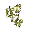
| Entry | Database: PDB / ID: 1jj7 | ||||||
|---|---|---|---|---|---|---|---|
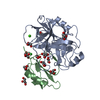
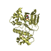
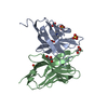
| Title | Crystal Structure of the C-terminal ATPase domain of human TAP1 | ||||||
 Components Components | PEPTIDE TRANSPORTER TAP1 | ||||||
 Keywords Keywords |  PROTEIN TRANSPORT / P-loop / ABC ATPase domain / helical domain PROTEIN TRANSPORT / P-loop / ABC ATPase domain / helical domain | ||||||
| Function / homology |  Function and homology information Function and homology informationABC-type antigen peptide transporter / ABC-type peptide antigen transporter activity / TAP complex / MHC class Ib protein binding / cytosol to endoplasmic reticulum transport / peptide transport / peptide transmembrane transporter activity / TAP1 binding / TAP2 binding / centriolar satellite ...ABC-type antigen peptide transporter / ABC-type peptide antigen transporter activity / TAP complex / MHC class Ib protein binding / cytosol to endoplasmic reticulum transport / peptide transport / peptide transmembrane transporter activity / TAP1 binding / TAP2 binding / centriolar satellite / MHC class I protein binding / endoplasmic reticulum-Golgi intermediate compartment membrane / ADP binding / Antigen Presentation: Folding, assembly and peptide loading of class I MHC / MHC class I peptide loading complex / transmembrane transport / antigen processing and presentation of endogenous peptide antigen via MHC class I / defense response / phagocytic vesicle membrane / peptide antigen binding / protein transport / ER-Phagosome pathway / adaptive immune response / endoplasmic reticulum membrane / endoplasmic reticulum / protein homodimerization activity / ATP hydrolysis activity / ATP binding / membrane / metal ion bindingSimilarity search - Function | ||||||
| Biological species |  Homo sapiens (human) Homo sapiens (human) | ||||||
| Method | X-RAY DIFFRACTION / SYNCHROTRON / MOLECULAR REPLACEMENT / Resolution: 2.4 Å | ||||||
 Authors Authors | Gaudet, R. / Wiley, D.C. | ||||||
 Citation Citation | Journal: EMBO J. / Year: 2001 Title: Structure of the ABC ATPase domain of human TAP1, the transporter associated with antigen processing. Authors: Gaudet, R. / Wiley, D.C. | ||||||
| History |
|
- Structure visualization
| Structure viewer | Molecule: MolmilJmol/JSmol |
|---|
- Downloads & links
-Download
| PDBx/mmCIF format | 1jj7.cif.gz | 65.9 KB | Display | PDBx/mmCIF format |
|---|---|---|---|---|
| PDB format | pdb1jj7.ent.gz | 46.9 KB | Display | PDB format |
| PDBx/mmJSON format | 1jj7.json.gz | Tree view | PDBx/mmJSON format | |
| Others |  Other downloads Other downloads |
-Validation report
| Arichive directory | https://data.pdbj.org/pub/pdb/validation_reports/jj/1jj7ftp://data.pdbj.org/pub/pdb/validation_reports/jj/1jj7 | HTTPS FTP |
|---|
-Related structure data
| Related structure data | 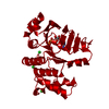 1b0uS S: Starting model for refinement |
|---|---|
| Similar structure data |


-Links
 PDBj
PDBj

- Assembly
| Deposited unit | 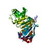
| ||||||||
|---|---|---|---|---|---|---|---|---|---|
| 1 |
| ||||||||
| Unit cell |
| ||||||||
| Details | The biological assembly is a monomer as contained in the asymmetric unit |
-Components
| #1: Protein | Mass: 28128.781 Da / Num. of mol.: 1 / Fragment: C-terminal ABC ATPase domain Source method: isolated from a genetically manipulated source Source: (gene. exp.) Homo sapiens (human) / Gene: TAP1 / Plasmid: pET15b / Species (production host): Escherichia coli / Production host:  Escherichia coli BL21(DE3) (bacteria) / Strain (production host): BL21(DE3) / References: UniProt: Q03518 Escherichia coli BL21(DE3) (bacteria) / Strain (production host): BL21(DE3) / References: UniProt: Q03518 |
|---|---|
| #2: Chemical | ChemComp-MG /   Mass: 24.305 Da / Num. of mol.: 1 / Source method: obtained synthetically / Formula: Mg Mass: 24.305 Da / Num. of mol.: 1 / Source method: obtained synthetically / Formula: Mg |
| #3: Chemical | ChemComp-ADP / Adenosine diphosphate  Mass: 427.201 Da / Num. of mol.: 1 / Source method: obtained synthetically / Formula: C10H15N5O10P2 / Comment: ADP, energy-carrying molecule*YM Mass: 427.201 Da / Num. of mol.: 1 / Source method: obtained synthetically / Formula: C10H15N5O10P2 / Comment: ADP, energy-carrying molecule*YM |
| #4: Water | ChemComp-HOH / Water Mass: 18.015 Da / Num. of mol.: 114 / Source method: isolated from a natural source / Formula: H2O Mass: 18.015 Da / Num. of mol.: 114 / Source method: isolated from a natural source / Formula: H2O |
-Experimental details
-Experiment
| Experiment | Method: X-RAY DIFFRACTION / Number of used crystals: 1 |
|---|
- Sample preparation
| Crystal | Density Matthews: 3.14 Å3/Da / Density % sol: 60.83 % | ||||||||||||||||||||||||||||||||||||||||||||||||||||||||||||||||||
|---|---|---|---|---|---|---|---|---|---|---|---|---|---|---|---|---|---|---|---|---|---|---|---|---|---|---|---|---|---|---|---|---|---|---|---|---|---|---|---|---|---|---|---|---|---|---|---|---|---|---|---|---|---|---|---|---|---|---|---|---|---|---|---|---|---|---|---|
| Crystal grow | Temperature: 277 K / Method: vapor diffusion, hanging drop / pH: 6.5 Details: 20% PEG8000, 100 mM sodium cacodylate, 200 mM magnesium acetate, pH 6.5, VAPOR DIFFUSION, HANGING DROP, temperature 277K | ||||||||||||||||||||||||||||||||||||||||||||||||||||||||||||||||||
| Crystal grow | *PLUS pH: 8 | ||||||||||||||||||||||||||||||||||||||||||||||||||||||||||||||||||
| Components of the solutions | *PLUS
|
-Data collection
| Diffraction | Mean temperature: 100 K |
|---|---|
| Diffraction source | Source: SYNCHROTRON / Site: APS  / Beamline: 14-BM-C / Wavelength: 1 Å / Beamline: 14-BM-C / Wavelength: 1 Å |
| Detector | Type: ADSC QUANTUM 4 / Detector: CCD / Date: Jun 27, 2000 |
| Radiation | Monochromator: SI 111 CHANNEL / Protocol: SINGLE WAVELENGTH / Monochromatic (M) / Laue (L): M / Scattering type: x-ray |
| Radiation wavelength | Wavelength: 1 Å / Relative weight: 1 |
| Reflection | Resolution: 2.4→30 Å / Num. all: 13714 / Num. obs: 13755 / % possible obs: 99.7 % / Observed criterion σ(F): -1000 / Observed criterion σ(I): -1000 / Redundancy: 3.7 % / Biso Wilson estimate: 52 Å2 / Rmerge(I) obs: 0.053 / Rsym value: 5.3 / Net I/σ(I): 15.9 |
| Reflection shell | Resolution: 2.4→2.49 Å / Redundancy: 3.7 % / Rmerge(I) obs: 0.401 / Mean I/σ(I) obs: 3.5 / Num. unique all: 1364 / Rsym value: 40.1 / % possible all: 100 |
| Reflection shell | *PLUS % possible obs: 100 % / Num. unique obs: 1364 |
- Processing
| Software |
| |||||||||||||||||||||||||
|---|---|---|---|---|---|---|---|---|---|---|---|---|---|---|---|---|---|---|---|---|---|---|---|---|---|---|
| Refinement | Method to determine structure: MOLECULAR REPLACEMENT Starting model: PDB ENTRY 1b0u Resolution: 2.4→30 Å / Isotropic thermal model: RESTRAINED / Cross valid method: THROUGHOUT / σ(F): 0 / Stereochemistry target values: Engh & Huber
| |||||||||||||||||||||||||
| Displacement parameters |
| |||||||||||||||||||||||||
| Refine analyze |
| |||||||||||||||||||||||||
| Refinement step | Cycle: LAST / Resolution: 2.4→30 Å
| |||||||||||||||||||||||||
| Refine LS restraints |
| |||||||||||||||||||||||||
| LS refinement shell | Resolution: 2.4→2.55 Å / Rfactor Rfree error: 0.02
| |||||||||||||||||||||||||
| Software | *PLUS Name: CNS / Version: 1 / Classification: refinement | |||||||||||||||||||||||||
| Refinement | *PLUS Lowest resolution: 30 Å / σ(F): 0 / Rfactor Rfree: 0.24 | |||||||||||||||||||||||||
| Solvent computation | *PLUS | |||||||||||||||||||||||||
| Displacement parameters | *PLUS | |||||||||||||||||||||||||
| Refine LS restraints | *PLUS
| |||||||||||||||||||||||||
| LS refinement shell | *PLUS Rfactor Rfree: 0.293 / Rfactor Rwork: 0.249 |
