 Movie
Movie Controller


+ Open data

- Basic information
| Entry | Database: PDB / ID: 1jdh | ||||||
|---|---|---|---|---|---|---|---|
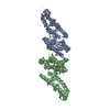
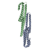
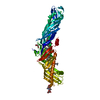
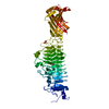
| Title | CRYSTAL STRUCTURE OF BETA-CATENIN AND HTCF-4 | ||||||
 Components Components |
| ||||||
 Keywords Keywords |  TRANSCRIPTION / BETA-CATENIN / TCF4 / PROTEIN-PROTEIN COMPLEX TRANSCRIPTION / BETA-CATENIN / TCF4 / PROTEIN-PROTEIN COMPLEX | ||||||
| Function / homology |  Function and homology information Function and homology informationcatenin-TCF7L2 complex / regulation of hormone metabolic process / negative regulation of type B pancreatic cell apoptotic process / Signaling by TCF7L2 mutants / Repression of WNT target genes / maintenance of DNA repeat elements / myoblast fate commitment / armadillo repeat domain binding / CDH11 homotypic and heterotypic interactions / Regulation of CDH19 Expression and Function ...catenin-TCF7L2 complex / regulation of hormone metabolic process / negative regulation of type B pancreatic cell apoptotic process / Signaling by TCF7L2 mutants / Repression of WNT target genes / maintenance of DNA repeat elements / myoblast fate commitment / armadillo repeat domain binding / CDH11 homotypic and heterotypic interactions / Regulation of CDH19 Expression and Function / positive regulation of heparan sulfate proteoglycan biosynthetic process / lung induction / positive regulation of branching involved in lung morphogenesis / cranial ganglion development / renal vesicle formation / renal inner medulla development / renal outer medulla development / nephron tubule formation / mesenchymal stem cell differentiation / beta-catenin-ICAT complex / metanephros morphogenesis / genitalia morphogenesis / embryonic skeletal limb joint morphogenesis / neural plate development / glial cell fate determination / regulation of secondary heart field cardioblast proliferation / astrocyte-dopaminergic neuron signaling / negative regulation of mitotic cell cycle, embryonic / canonical Wnt signaling pathway involved in mesenchymal stem cell differentiation / oviduct development / beta-catenin-TCF7L2 complex / regulation of nephron tubule epithelial cell differentiation / negative regulation of mesenchymal to epithelial transition involved in metanephros morphogenesis / regulation of timing of anagen / Binding of TCF/LEF:CTNNB1 to target gene promoters / central nervous system vasculogenesis / RUNX3 regulates WNT signaling / regulation of centriole-centriole cohesion / Regulation of CDH11 function / regulation of centromeric sister chromatid cohesion / embryonic axis specification / endodermal cell fate commitment / positive regulation of fibroblast growth factor receptor signaling pathway / regulation of fibroblast proliferation / Scrib-APC-beta-catenin complex / beta-catenin-TCF complex / lens morphogenesis in camera-type eye / dorsal root ganglion development / synaptic vesicle clustering / gamma-catenin binding / acinar cell differentiation / dorsal/ventral axis specification / proximal/distal pattern formation / neuron fate determination / layer formation in cerebral cortex / positive regulation of myoblast proliferation / positive regulation of endothelial cell differentiation / sympathetic ganglion development / establishment of blood-retinal barrier / fungiform papilla formation / lung epithelial cell differentiation / embryonic foregut morphogenesis / hindbrain development / regulation of calcium ion import / positive regulation of determination of dorsal identity / positive regulation of skeletal muscle tissue development / ectoderm development / positive regulation of odontoblast differentiation / cranial skeletal system development / endothelial tube morphogenesis / regulation of protein localization to cell surface / hair cell differentiation / cellular response to indole-3-methanol / mesenchymal cell proliferation involved in lung development / presynaptic active zone cytoplasmic component / detection of muscle stretch / smooth muscle cell differentiation / histone methyltransferase binding / midbrain dopaminergic neuron differentiation / alpha-catenin binding / flotillin complex / establishment of blood-brain barrier / Germ layer formation at gastrulation / male genitalia development / negative regulation of oligodendrocyte differentiation / fascia adherens / epithelial cell proliferation involved in prostate gland development / apicolateral plasma membrane / embryonic brain development / Formation of definitive endoderm / epithelial cell differentiation involved in prostate gland development / positive regulation of epithelial cell proliferation involved in prostate gland development / regulation of smooth muscle cell proliferation / negative regulation of oxidative stress-induced neuron intrinsic apoptotic signaling pathway / oocyte development / beta-catenin destruction complex / lung-associated mesenchyme development / Formation of axial mesoderm / negative regulation of protein sumoylation / adherens junction assemblySimilarity search - Function | ||||||
| Biological species |  Homo sapiens (human) Homo sapiens (human) | ||||||
| Method | X-RAY DIFFRACTION / SYNCHROTRON / MOLECULAR REPLACEMENT / Resolution: 1.9 Å | ||||||
 Authors Authors | Graham, T.A. / Ferkey, D.M. / Mao, F. / Kimelman, D. / Xu, W. | ||||||
 Citation Citation | Journal: Nat.Struct.Biol. / Year: 2001 Title: Tcf4 can specifically recognize beta-catenin using alternative conformations. Authors: Graham, T.A. / Ferkey, D.M. / Mao, F. / Kimelman, D. / Xu, W. | ||||||
| History |
|
- Structure visualization
| Structure viewer | Molecule: MolmilJmol/JSmol |
|---|
- Downloads & links
-Download
| PDBx/mmCIF format | 1jdh.cif.gz | 121.8 KB | Display | PDBx/mmCIF format |
|---|---|---|---|---|
| PDB format | pdb1jdh.ent.gz | 91.1 KB | Display | PDB format |
| PDBx/mmJSON format | 1jdh.json.gz | Tree view | PDBx/mmJSON format | |
| Others |  Other downloads Other downloads |
-Validation report
| Arichive directory | https://data.pdbj.org/pub/pdb/validation_reports/jd/1jdhftp://data.pdbj.org/pub/pdb/validation_reports/jd/1jdh | HTTPS FTP |
|---|
-Related structure data
| Similar structure data |
|---|


-Links
 PDBj
PDBj


- Assembly
| Deposited unit | 
| ||||||||
|---|---|---|---|---|---|---|---|---|---|
| 1 |
| ||||||||
| Unit cell |
|
-Components
| #1: Protein | Catenin beta-1 Mass: 57699.902 Da / Num. of mol.: 1 / Fragment: Residues 135-663 Source method: isolated from a genetically manipulated source Source: (gene. exp.) Homo sapiens (human) / Production host:  Escherichia coli (E. coli) / References: UniProt: P35222 Escherichia coli (E. coli) / References: UniProt: P35222 |
|---|---|
| #2: Protein/peptide | Mass: 4131.336 Da / Num. of mol.: 1 / Fragment: Residues 12-49 Source method: isolated from a genetically manipulated source Source: (gene. exp.) Homo sapiens (human) / Production host: Escherichia coli (E. coli) / References: UniProt: Q9NQB0 |
| #3: Water | ChemComp-HOH / Water Mass: 18.015 Da / Num. of mol.: 393 / Source method: isolated from a natural source / Formula: H2O Mass: 18.015 Da / Num. of mol.: 393 / Source method: isolated from a natural source / Formula: H2O |
-Experimental details
-Experiment
| Experiment | Method: X-RAY DIFFRACTION / Number of used crystals: 1 |
|---|
- Sample preparation
| Crystal | Density Matthews: 2.43 Å3/Da / Density % sol: 49.47 % | ||||||||||||||||||||||||||||||||||||||||||||||||||||||||
|---|---|---|---|---|---|---|---|---|---|---|---|---|---|---|---|---|---|---|---|---|---|---|---|---|---|---|---|---|---|---|---|---|---|---|---|---|---|---|---|---|---|---|---|---|---|---|---|---|---|---|---|---|---|---|---|---|---|
| Crystal grow | *PLUS pH: 5.8 / Method: vapor diffusion, hanging drop | ||||||||||||||||||||||||||||||||||||||||||||||||||||||||
| Components of the solutions | *PLUS
|
-Data collection
| Diffraction source | Source: SYNCHROTRON / Site: APS  / Type: APS / Wavelength: 1.063 / Type: APS / Wavelength: 1.063 |
|---|---|
| Radiation | Protocol: SINGLE WAVELENGTH / Monochromatic (M) / Laue (L): M / Scattering type: x-ray |
| Radiation wavelength | Wavelength: 1.063 Å / Relative weight: 1 |
| Reflection | Resolution: 1.9→20 Å / Num. obs: 47949 / % possible obs: 98.8 % / Redundancy: 9.98 % / Biso Wilson estimate: 23 Å2 / Rsym value: 0.76 / Net I/σ(I): 23.9 |
| Reflection shell | Resolution: 1.9→1.97 Å / Rsym value: 0.306 |
- Processing
| Software |
| ||||||||||||||||||||||||||||||||||||||||||||||||||||||||||||
|---|---|---|---|---|---|---|---|---|---|---|---|---|---|---|---|---|---|---|---|---|---|---|---|---|---|---|---|---|---|---|---|---|---|---|---|---|---|---|---|---|---|---|---|---|---|---|---|---|---|---|---|---|---|---|---|---|---|---|---|---|---|
| Refinement | Method to determine structure: MOLECULAR REPLACEMENT / Resolution: 1.9→20 Å / Rfactor Rfree error: 0.004 / Data cutoff high absF: 727211.15 / Data cutoff low absF: 0 / Isotropic thermal model: RESTRAINED / Cross valid method: THROUGHOUT / σ(F): 2 / Details: BULK SOLVENT MODEL USED
| ||||||||||||||||||||||||||||||||||||||||||||||||||||||||||||
| Solvent computation | Solvent model: FLAT MODEL / Bsol: 52.16 Å2 / ksol: 0.345 e/Å3 | ||||||||||||||||||||||||||||||||||||||||||||||||||||||||||||
| Displacement parameters | Biso mean: 39 Å2
| ||||||||||||||||||||||||||||||||||||||||||||||||||||||||||||
| Refine analyze |
| ||||||||||||||||||||||||||||||||||||||||||||||||||||||||||||
| Refinement step | Cycle: LAST / Resolution: 1.9→20 Å
| ||||||||||||||||||||||||||||||||||||||||||||||||||||||||||||
| Refine LS restraints |
| ||||||||||||||||||||||||||||||||||||||||||||||||||||||||||||
| LS refinement shell | Resolution: 1.9→2.02 Å / Rfactor Rfree error: 0.021 / Total num. of bins used: 6
| ||||||||||||||||||||||||||||||||||||||||||||||||||||||||||||
| Xplor file |
| ||||||||||||||||||||||||||||||||||||||||||||||||||||||||||||
| Software | *PLUS Name: CNS / Version: 0.5 / Classification: refinement | ||||||||||||||||||||||||||||||||||||||||||||||||||||||||||||
| Refinement | *PLUS Num. reflection obs: 36861 / σ(F): 2 / Num. reflection Rfree: 3719 / % reflection Rfree: 9.7 % / Rfactor obs: 0.202 | ||||||||||||||||||||||||||||||||||||||||||||||||||||||||||||
| Solvent computation | *PLUS | ||||||||||||||||||||||||||||||||||||||||||||||||||||||||||||
| Displacement parameters | *PLUS Biso mean: 39 Å2 | ||||||||||||||||||||||||||||||||||||||||||||||||||||||||||||
| Refine LS restraints | *PLUS
| ||||||||||||||||||||||||||||||||||||||||||||||||||||||||||||
| LS refinement shell | *PLUS Rfactor Rfree: 0.262 / % reflection Rfree: 5 % / Rfactor Rwork: 0.221 |
