 Movie
Movie Controller


+ Open data

- Basic information
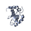
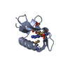
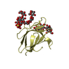
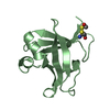
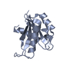
| Entry | Database: PDB / ID: 1jd6 | ||||||
|---|---|---|---|---|---|---|---|
| Title | Crystal Structure of DIAP1-BIR2/Hid Complex | ||||||
 Components Components |
| ||||||
 Keywords Keywords | Hydrolase/Peptide /  APOPTOSIS / IAP / HID / CASPASE ACTIVATION / DROSOPHILA / Hydrolase-Peptide complex APOPTOSIS / IAP / HID / CASPASE ACTIVATION / DROSOPHILA / Hydrolase-Peptide complex | ||||||
| Function / homology |  Function and homology information Function and homology information: / instar larval or pupal development / positive regulation of cellular response to X-ray / BIR domain binding / larval midgut cell programmed cell death / SMAC, XIAP-regulated apoptotic response / DS ligand bound to FT receptor / response to red light / head involution / negative regulation of compound eye retinal cell programmed cell death ...: / instar larval or pupal development / positive regulation of cellular response to X-ray / BIR domain binding / larval midgut cell programmed cell death / SMAC, XIAP-regulated apoptotic response / DS ligand bound to FT receptor / response to red light / head involution / negative regulation of compound eye retinal cell programmed cell death / antennal morphogenesis / positive regulation of apoptotic process involved in development / Deactivation of the beta-catenin transactivating complex / Regulation of necroptotic cell death / Regulation of PTEN localization / sensory organ precursor cell division / Regulation of PTEN stability and activity / open tracheal system development / regulation of cysteine-type endopeptidase activity involved in apoptotic process / spermatid nucleus differentiation / positive regulation of Toll signaling pathway / border follicle cell migration / : / positive regulation of border follicle cell migration / chaeta development / cell death / sex differentiation / caspase binding / positive regulation of cysteine-type endopeptidase activity involved in execution phase of apoptosis / programmed cell death / negative regulation of JNK cascade / regulation of organ growth / positive regulation of cysteine-type endopeptidase activity / protein neddylation / ubiquitin conjugating enzyme binding / ubiquitin-like protein conjugating enzyme binding / dendrite morphogenesis / NEDD8 ligase activity / cysteine-type endopeptidase inhibitor activity / ubiquitin-specific protease binding / entrainment of circadian clock by photoperiod / cysteine-type endopeptidase inhibitor activity involved in apoptotic process / positive regulation of macroautophagy / protein K48-linked ubiquitination / protein autoubiquitination / cellular response to starvation / positive regulation of protein ubiquitination / apoptotic signaling pathway / RING-type E3 ubiquitin transferase / cellular response to gamma radiation / Wnt signaling pathway / protein polyubiquitination / ubiquitin-protein transferase activity / positive regulation of canonical Wnt signaling pathway / ubiquitin protein ligase activity / intrinsic apoptotic signaling pathway in response to DNA damage / spermatogenesis / regulation of cell cycle / positive regulation of apoptotic process / apoptotic process / ubiquitin protein ligase binding / negative regulation of apoptotic process / perinuclear region of cytoplasm / mitochondrion / zinc ion binding / nucleus / plasma membrane / cytosol / cytoplasmSimilarity search - Function | ||||||
| Biological species |  Drosophila melanogaster (fruit fly) Drosophila melanogaster (fruit fly) | ||||||
| Method | X-RAY DIFFRACTION / MOLECULAR REPLACEMENT / Resolution: 2.7 Å | ||||||
 Authors Authors | Wu, J.W. / Cocina, A.E. / Chai, J. / Hay, B.A. / Shi, Y. | ||||||
 Citation Citation | Journal: Mol.Cell / Year: 2001 Title: Structural analysis of a functional DIAP1 fragment bound to grim and hid peptides. Authors: Wu, J.W. / Cocina, A.E. / Chai, J. / Hay, B.A. / Shi, Y. | ||||||
| History |
|
- Structure visualization
| Structure viewer | Molecule: MolmilJmol/JSmol |
|---|
- Downloads & links
-Download
| PDBx/mmCIF format | 1jd6.cif.gz | 35.3 KB | Display | PDBx/mmCIF format |
|---|---|---|---|---|
| PDB format | pdb1jd6.ent.gz | 23.6 KB | Display | PDB format |
| PDBx/mmJSON format | 1jd6.json.gz | Tree view | PDBx/mmJSON format | |
| Others |  Other downloads Other downloads |
-Validation report
| Arichive directory | https://data.pdbj.org/pub/pdb/validation_reports/jd/1jd6ftp://data.pdbj.org/pub/pdb/validation_reports/jd/1jd6 | HTTPS FTP |
|---|
-Related structure data


-Links
 PDBj
PDBj


- Assembly
| Deposited unit | 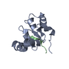
| ||||||||
|---|---|---|---|---|---|---|---|---|---|
| 1 |
| ||||||||
| Unit cell |
|
-Components
| #1: Protein | Mass: 14078.695 Da / Num. of mol.: 1 Source method: isolated from a genetically manipulated source Source: (gene. exp.) Drosophila melanogaster (fruit fly) / Gene: DIAP1 / Plasmid: PGEX2T / Species (production host): Escherichia coli / Production host:  Escherichia coli BL21(DE3) (bacteria) / Strain (production host): BL21(DE3) / References: UniProt: Q24306 Escherichia coli BL21(DE3) (bacteria) / Strain (production host): BL21(DE3) / References: UniProt: Q24306 |
|---|---|
| #2: Protein/peptide | Mass: 1049.176 Da / Num. of mol.: 1 / Source method: obtained synthetically Details: This peptide was chemically synthesized. The sequence is naturally found in Drosophila melanogaster (fruit fly). References: GenBank: Q24106, UniProt: Q24106*PLUS |
| #3: Chemical | ChemComp-ZN /   Mass: 65.409 Da / Num. of mol.: 1 / Source method: obtained synthetically / Formula: Zn Mass: 65.409 Da / Num. of mol.: 1 / Source method: obtained synthetically / Formula: Zn |
-Experimental details
-Experiment
| Experiment | Method: X-RAY DIFFRACTION / Number of used crystals: 1 |
|---|
- Sample preparation
| Crystal | Density Matthews: 2.45 Å3/Da / Density % sol: 49.79 % | ||||||||||||||||||||||||||||||
|---|---|---|---|---|---|---|---|---|---|---|---|---|---|---|---|---|---|---|---|---|---|---|---|---|---|---|---|---|---|---|---|
| Crystal grow | Temperature: 296 K / Method: vapor diffusion, hanging drop / pH: 8.5 Details: TRIS, AMMONIUM DIHYDROGEN PHOSPHATE, pH 8.5, VAPOR DIFFUSION, HANGING DROP, temperature 296K | ||||||||||||||||||||||||||||||
| Crystal grow | *PLUS | ||||||||||||||||||||||||||||||
| Components of the solutions | *PLUS
|
-Data collection
| Diffraction | Mean temperature: 100 K |
|---|---|
| Diffraction source | Source: ROTATING ANODE / Type: RIGAKU RU200 / Wavelength: 1.5418 Å |
| Detector | Type: RIGAKU RAXIS IV / Detector: IMAGE PLATE / Date: Jan 8, 2001 / Details: MIRRORS |
| Radiation | Monochromator: GRAPHITE / Protocol: SINGLE WAVELENGTH / Monochromatic (M) / Laue (L): M / Scattering type: x-ray |
| Radiation wavelength | Wavelength: 1.5418 Å / Relative weight: 1 |
| Reflection | Resolution: 2.7→99 Å / Num. all: 4613 / Num. obs: 4535 / % possible obs: 98.3 % / Observed criterion σ(F): 1.4 / Observed criterion σ(I): 2 / Redundancy: 19 % / Biso Wilson estimate: 36 Å2 / Rmerge(I) obs: 0.133 / Net I/σ(I): 25 |
| Reflection shell | Resolution: 2.7→2.8 Å / Redundancy: 12 % / Rmerge(I) obs: 0.043 |
| Reflection | *PLUS Lowest resolution: 99 Å / Num. measured all: 87781 |
| Reflection shell | *PLUS % possible obs: 98.4 % / Rmerge(I) obs: 0.381 |
- Processing
| Software |
| ||||||||||||||||||||
|---|---|---|---|---|---|---|---|---|---|---|---|---|---|---|---|---|---|---|---|---|---|
| Refinement | Method to determine structure: MOLECULAR REPLACEMENT / Resolution: 2.7→20 Å / Cross valid method: THROUGHOUT / σ(F): 0 / σ(I): 0 / Stereochemistry target values: Engh & Huber
| ||||||||||||||||||||
| Refinement step | Cycle: LAST / Resolution: 2.7→20 Å
| ||||||||||||||||||||
| Refine LS restraints |
| ||||||||||||||||||||
| LS refinement shell | Resolution: 2.7→2.82 Å / Rfactor Rfree error: 0.012
|
