 Movie
Movie Controller

[English] 日本語
 Yorodumi
- PDB-1jc7: The Laminin-Binding Domain of Agrin is Structurally Related to N-... -
+ Open data

- Basic information
| Entry | Database: PDB / ID: 1jc7 | ||||||
|---|---|---|---|---|---|---|---|
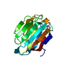
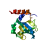
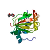
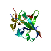
| Title | The Laminin-Binding Domain of Agrin is Structurally Related to N-TIMP-1 | ||||||
 Components Components | Agrin | ||||||
 Keywords Keywords | CELL ADHESION / NEUROMUSCULAR JUNCTION / AGRIN / INTERACTION COILED-DOIL PROTEINS WITH GLOBULAR PROTEINS / OB-FOLD / TIMP | ||||||
| Function / homology |  Function and homology information Function and homology informationextracellular matrix of synaptic cleft / acetylcholine receptor regulator activity / positive regulation of synaptic assembly at neuromuscular junction / skeletal muscle acetylcholine-gated channel clustering / chondroitin sulfate binding / laminin-1 binding / sialic acid binding / neuron cell-cell adhesion / dystroglycan binding / basal part of cell ...extracellular matrix of synaptic cleft / acetylcholine receptor regulator activity / positive regulation of synaptic assembly at neuromuscular junction / skeletal muscle acetylcholine-gated channel clustering / chondroitin sulfate binding / laminin-1 binding / sialic acid binding / neuron cell-cell adhesion / dystroglycan binding / basal part of cell / photoreceptor ribbon synapse / filopodium assembly / glycosaminoglycan binding / extracellular matrix binding / heparan sulfate proteoglycan binding / positive regulation of filopodium assembly / neuromuscular junction development / transmembrane receptor protein tyrosine kinase activator activity / receptor clustering / basement membrane / laminin binding / extracellular matrix / brain development / neuromuscular junction / G protein-coupled acetylcholine receptor signaling pathway / positive regulation of GTPase activity / negative regulation of neuron projection development / signaling receptor activity / heparin binding / nervous system development / actin cytoskeleton organization / cell differentiation / membrane raft / axon / glutamatergic synapse / synapse / calcium ion binding / positive regulation of transcription by RNA polymerase II / extracellular space / extracellular region / plasma membraneSimilarity search - Function | ||||||
| Biological species |  Gallus gallus (chicken) Gallus gallus (chicken) | ||||||
| Method | X-RAY DIFFRACTION / SYNCHROTRON / Resolution: 2.73 Å | ||||||
 Authors Authors | Stetefeld, J. | ||||||
 Citation Citation | Journal: Nat.Struct.Biol. / Year: 2001 Title: The laminin-binding domain of agrin is structurally related to N-TIMP-1. Authors: Stetefeld, J. / Jenny, M. / Schulthess, T. / Landwehr, R. / Schumacher, B. / Frank, S. / Ruegg, M.A. / Engel, J. / Kammerer, R.A. | ||||||
| History |
|
- Structure visualization
| Structure viewer | Molecule: MolmilJmol/JSmol |
|---|
- Downloads & links
-Download
| PDBx/mmCIF format | 1jc7.cif.gz | 33.8 KB | Display | PDBx/mmCIF format |
|---|---|---|---|---|
| PDB format | pdb1jc7.ent.gz | 25.6 KB | Display | PDB format |
| PDBx/mmJSON format | 1jc7.json.gz | Tree view | PDBx/mmJSON format | |
| Others |  Other downloads Other downloads |
-Validation report
| Arichive directory | https://data.pdbj.org/pub/pdb/validation_reports/jc/1jc7ftp://data.pdbj.org/pub/pdb/validation_reports/jc/1jc7 | HTTPS FTP |
|---|
-Related structure data


-Links
 PDBj
PDBj


- Assembly
| Deposited unit | 
| ||||||||
|---|---|---|---|---|---|---|---|---|---|
| 1 |
| ||||||||
| Unit cell |
|
-Components
| #1: Protein | Mass: 15147.343 Da / Num. of mol.: 1 / Fragment: LAMININ-BINDING DOMAIN Source method: isolated from a genetically manipulated source Source: (gene. exp.) Gallus gallus (chicken) / Production host:  Escherichia coli (E. coli) / References: UniProt: Q90685, UniProt: P31696*PLUS Escherichia coli (E. coli) / References: UniProt: Q90685, UniProt: P31696*PLUS |
|---|---|
| #2: Chemical | ChemComp-CL / Chloride  Mass: 35.453 Da / Num. of mol.: 1 / Source method: obtained synthetically / Formula: Cl Mass: 35.453 Da / Num. of mol.: 1 / Source method: obtained synthetically / Formula: Cl |
-Experimental details
-Experiment
| Experiment | Method: X-RAY DIFFRACTION / Number of used crystals: 1 |
|---|
- Sample preparation
| Crystal | Density Matthews: 3.36 Å3/Da / Density % sol: 63.34 % | |||||||||||||||||||||||||||||||||||
|---|---|---|---|---|---|---|---|---|---|---|---|---|---|---|---|---|---|---|---|---|---|---|---|---|---|---|---|---|---|---|---|---|---|---|---|---|
| Crystal grow | Temperature: 298 K / Method: vapor diffusion, hanging drop / pH: 5.8 Details: PEG4000, pH 5.8, VAPOR DIFFUSION, HANGING DROP, temperature 298K | |||||||||||||||||||||||||||||||||||
| Crystal grow | *PLUS Temperature: 4 ℃ | |||||||||||||||||||||||||||||||||||
| Components of the solutions | *PLUS
|
-Data collection
| Diffraction source | Source: SYNCHROTRON / Site: EMBL/DESY, Hamburg  / Beamline: X11 / Wavelength: 0.847 Å / Beamline: X11 / Wavelength: 0.847 Å |
|---|---|
| Detector | Type: MARRESEARCH / Detector: IMAGE PLATE |
| Radiation | Protocol: SINGLE WAVELENGTH / Monochromatic (M) / Laue (L): M / Scattering type: x-ray |
| Radiation wavelength | Wavelength: 0.847 Å / Relative weight: 1 |
| Reflection | Biso Wilson estimate: 26.4 Å2 |
| Reflection | *PLUS Highest resolution: 2.7 Å / % possible obs: 99.1 % / Redundancy: 4.5 % / Rmerge(I) obs: 0.086 |
| Reflection shell | *PLUS % possible obs: 94.3 % / Rmerge(I) obs: 0.272 |
- Processing
| Software |
| ||||||||||||||||||||||||||||||||||||||||||||||||||||||||||||
|---|---|---|---|---|---|---|---|---|---|---|---|---|---|---|---|---|---|---|---|---|---|---|---|---|---|---|---|---|---|---|---|---|---|---|---|---|---|---|---|---|---|---|---|---|---|---|---|---|---|---|---|---|---|---|---|---|---|---|---|---|---|
| Refinement | Resolution: 2.73→27.92 Å / Rfactor Rfree error: 0.011 / Data cutoff high absF: 664563.5 / Data cutoff low absF: 0 / Isotropic thermal model: RESTRAINED / Cross valid method: THROUGHOUT
| ||||||||||||||||||||||||||||||||||||||||||||||||||||||||||||
| Solvent computation | Solvent model: FLAT MODEL / Bsol: 24.06 Å2 / ksol: 0.304 e/Å3 | ||||||||||||||||||||||||||||||||||||||||||||||||||||||||||||
| Displacement parameters | Biso mean: 35.9 Å2
| ||||||||||||||||||||||||||||||||||||||||||||||||||||||||||||
| Refine analyze |
| ||||||||||||||||||||||||||||||||||||||||||||||||||||||||||||
| Refinement step | Cycle: LAST / Resolution: 2.73→27.92 Å
| ||||||||||||||||||||||||||||||||||||||||||||||||||||||||||||
| Refine LS restraints |
| ||||||||||||||||||||||||||||||||||||||||||||||||||||||||||||
| LS refinement shell | Resolution: 2.7→2.87 Å / Rfactor Rfree error: 0.05 / Total num. of bins used: 6
| ||||||||||||||||||||||||||||||||||||||||||||||||||||||||||||
| Xplor file |
| ||||||||||||||||||||||||||||||||||||||||||||||||||||||||||||
| Software | *PLUS Name: CNS / Version: 1 / Classification: refinement | ||||||||||||||||||||||||||||||||||||||||||||||||||||||||||||
| Refinement | *PLUS % reflection Rfree: 10.4 % | ||||||||||||||||||||||||||||||||||||||||||||||||||||||||||||
| Solvent computation | *PLUS | ||||||||||||||||||||||||||||||||||||||||||||||||||||||||||||
| Displacement parameters | *PLUS Biso mean: 35.9 Å2 | ||||||||||||||||||||||||||||||||||||||||||||||||||||||||||||
| Refine LS restraints | *PLUS
| ||||||||||||||||||||||||||||||||||||||||||||||||||||||||||||
| LS refinement shell | *PLUS Rfactor Rfree: 0.342 / % reflection Rfree: 9.3 % / Rfactor Rwork: 0.316 |
