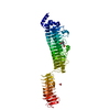
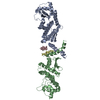
- PDB-1jad: C-terminal Domain of Turkey PLC-beta -
+
Open data
ID or keywords:
Loading...
-
Basic information
Entry
Database: PDB / ID: 1jad
Title
C-terminal Domain of Turkey PLC-beta
Components
phospholipase C beta
Keywords
HYDROLASE / AlPHA HELICAL COILED COIL
Function / homology
Function and homology information
phosphoinositide phospholipase C / phosphatidylinositol phospholipase C activity / lipid catabolic process / intracellular signal transduction / calcium ion binding / cytosol Similarity search - Function
Phospholipase C Beta; Chain: A / Phospholipase C beta, distal C-terminal domain / 1-phosphatidylinositol 4,5-bisphosphate phosphodiesterase beta-2 / Phospholipase C-beta, C-terminal domain / PLC-beta C terminal / PLC-beta, PH domain / Phospholipase C-beta, C-terminal domain superfamily / PH domain / Phosphatidylinositol-4, 5-bisphosphate phosphodiesterase beta / Phosphoinositide-specific phospholipase C, EF-hand-like domain ...Phospholipase C Beta; Chain: A / Phospholipase C beta, distal C-terminal domain / 1-phosphatidylinositol 4,5-bisphosphate phosphodiesterase beta-2 / Phospholipase C-beta, C-terminal domain / PLC-beta C terminal / PLC-beta, PH domain / Phospholipase C-beta, C-terminal domain superfamily / PH domain / Phosphatidylinositol-4, 5-bisphosphate phosphodiesterase beta / Phosphoinositide-specific phospholipase C, EF-hand-like domain / Phosphoinositide-specific phospholipase C, efhand-like / Phosphoinositide phospholipase C family / Phospholipase C, phosphatidylinositol-specific, Y domain / Phosphatidylinositol-specific phospholipase C, Y domain / Phosphatidylinositol-specific phospholipase Y-box domain profile. / Phospholipase C, catalytic domain (part); domain Y / Phosphatidylinositol-specific phospholipase C, X domain / Phosphatidylinositol-specific phospholipase C, X domain / Phospholipase C, catalytic domain (part); domain X / PLC-like phosphodiesterase, TIM beta/alpha-barrel domain superfamily / C2 domain / Protein kinase C conserved region 2 (CalB) / C2 domain / C2 domain profile. / C2 domain superfamily / EF-hand domain pair / Up-down Bundle / Mainly Alpha Similarity search - Domain/homology
In the structure databanks used in Yorodumi, some data are registered as the other names, "COVID-19 virus" and "2019-nCoV". Here are the details of the virus and the list of structure data.
Jan 31, 2019. EMDB accession codes are about to change! (news from PDBe EMDB page)
EMDB accession codes are about to change! (news from PDBe EMDB page)
The allocation of 4 digits for EMDB accession codes will soon come to an end. Whilst these codes will remain in use, new EMDB accession codes will include an additional digit and will expand incrementally as the available range of codes is exhausted. The current 4-digit format prefixed with “EMD-” (i.e. EMD-XXXX) will advance to a 5-digit format (i.e. EMD-XXXXX), and so on. It is currently estimated that the 4-digit codes will be depleted around Spring 2019, at which point the 5-digit format will come into force.
The EM Navigator/Yorodumi systems omit the EMD- prefix.
Related info.:Q: What is EMD? / ID/Accession-code notation in Yorodumi/EM Navigator
Yorodumi is a browser for structure data from EMDB, PDB, SASBDB, etc.
This page is also the successor to EM Navigator detail page, and also detail information page/front-end page for Omokage search.
The word "yorodu" (or yorozu) is an old Japanese word meaning "ten thousand". "mi" (miru) is to see.
Related info.:EMDB / PDB / SASBDB / Comparison of 3 databanks / Yorodumi Search / Aug 31, 2016. New EM Navigator & Yorodumi / Yorodumi Papers / Jmol/JSmol / Function and homology information / Changes in new EM Navigator and Yorodumi
 Movie
Movie Controller


 Open data
 Basic information
 Components
 Keywords
 HYDROLASE / AlPHA HELICAL COILED COIL
 Function and homology information
 Authors
 Citation
 Structure visualization
 Downloads & links
 Other downloads
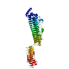


 PDBj
PDBj


 Assembly


 Mass: 96.063 Da / Num. of mol.: 1 / Source method: obtained synthetically / Formula: SO4
 Mass: 18.015 Da / Num. of mol.: 143 / Source method: isolated from a natural source / Formula: H2O
 Sample preparation
 / Beamline: BL1-5 / Wavelength: 0.97949, 0.97940, 0.97526
 Processing