 Movie
Movie Controller


+ Open data

- Basic information
| Entry | Database: PDB / ID: 1j90 | ||||||
|---|---|---|---|---|---|---|---|
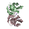
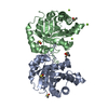
| Title | Crystal Structure of Drosophila Deoxyribonucleoside Kinase | ||||||
 Components Components | Deoxyribonucleoside kinase | ||||||
 Keywords Keywords |  TRANSFERASE / Protein-deoxynucleoside complex TRANSFERASE / Protein-deoxynucleoside complex | ||||||
| Function / homology |  Function and homology informationdeoxynucleoside kinase / Pyrimidine salvage / deoxynucleoside kinase activity / nucleoside salvage / uridine kinase activity / deoxycytidine kinase activity / nucleoside phosphate biosynthetic process / thymidine kinase activity / deoxyguanosine kinase activity / deoxyadenosine kinase activity ...deoxynucleoside kinase / Pyrimidine salvage / deoxynucleoside kinase activity / nucleoside salvage / uridine kinase activity / deoxycytidine kinase activity / nucleoside phosphate biosynthetic process / thymidine kinase activity / deoxyguanosine kinase activity / deoxyadenosine kinase activity / cytidine kinase activity / DNA biosynthetic process / kinase activity / phosphorylation / mitochondrion / ATP binding / cytoplasm Function and homology informationdeoxynucleoside kinase / Pyrimidine salvage / deoxynucleoside kinase activity / nucleoside salvage / uridine kinase activity / deoxycytidine kinase activity / nucleoside phosphate biosynthetic process / thymidine kinase activity / deoxyguanosine kinase activity / deoxyadenosine kinase activity ...deoxynucleoside kinase / Pyrimidine salvage / deoxynucleoside kinase activity / nucleoside salvage / uridine kinase activity / deoxycytidine kinase activity / nucleoside phosphate biosynthetic process / thymidine kinase activity / deoxyguanosine kinase activity / deoxyadenosine kinase activity / cytidine kinase activity / DNA biosynthetic process / kinase activity / phosphorylation / mitochondrion / ATP binding / cytoplasmSimilarity search - Function | ||||||
| Biological species |  Drosophila melanogaster (fruit fly) Drosophila melanogaster (fruit fly) | ||||||
| Method | X-RAY DIFFRACTION / SYNCHROTRON / MAD / Resolution: 2.56 Å | ||||||
 Authors Authors | Johansson, K. / Ramaswamy, S. / Ljungkrantz, C. / Knecht, W. / Piskur, J. / Munch-Petersen, B. / Eriksson, S. / Eklund, H. | ||||||
 Citation Citation | Journal: Nat.Struct.Biol. / Year: 2001 Title: Structural basis for substrate specificities of cellular deoxyribonucleoside kinases. Authors: Johansson, K. / Ramaswamy, S. / Ljungcrantz, C. / Knecht, W. / Piskur, J. / Munch-Petersen, B. / Eriksson, S. / Eklund, H. | ||||||
| History |
| ||||||
| Remark 999 | SEQUENCE THE LAST 20 RESIDUES OF THE SEQUENCE DATABASE REFERENCE, RESIDUES 231-250, WERE TRUNCATED. |
- Structure visualization
| Structure viewer | Molecule: MolmilJmol/JSmol |
|---|
- Downloads & links
-Download
| PDBx/mmCIF format | 1j90.cif.gz | 94.7 KB | Display | PDBx/mmCIF format |
|---|---|---|---|---|
| PDB format | pdb1j90.ent.gz | 72.2 KB | Display | PDB format |
| PDBx/mmJSON format | 1j90.json.gz | Tree view | PDBx/mmJSON format | |
| Others |  Other downloads Other downloads |
-Validation report
| Arichive directory | https://data.pdbj.org/pub/pdb/validation_reports/j9/1j90ftp://data.pdbj.org/pub/pdb/validation_reports/j9/1j90 | HTTPS FTP |
|---|
-Related structure data


-Links
 PDBj
PDBj- Assembly
| Deposited unit | 
| ||||||||
|---|---|---|---|---|---|---|---|---|---|
| 1 |
| ||||||||
| Unit cell |
| ||||||||
| Details | The biological dimer is present in the asymmetric unit |
-Components
| #1: Protein | Mass: 26906.707 Da / Num. of mol.: 2 / Fragment: Truncation mutant / Mutation: Deletion of last 20 residues Source method: isolated from a genetically manipulated source Source: (gene. exp.) Drosophila melanogaster (fruit fly) / Production host:  Escherichia coli (E. coli) / References: UniProt: Q9XZT6 Escherichia coli (E. coli) / References: UniProt: Q9XZT6#2: Chemical | Sulfate  Mass: 96.063 Da / Num. of mol.: 2 / Source method: obtained synthetically / Formula: SO4 Mass: 96.063 Da / Num. of mol.: 2 / Source method: obtained synthetically / Formula: SO4#3: Chemical | Deoxycytidine  Type: DNA OH 5 prime terminus / Mass: 227.217 Da / Num. of mol.: 2 / Source method: obtained synthetically / Formula: C9H13N3O4 Type: DNA OH 5 prime terminus / Mass: 227.217 Da / Num. of mol.: 2 / Source method: obtained synthetically / Formula: C9H13N3O4#4: Water | ChemComp-HOH / | Water Mass: 18.015 Da / Num. of mol.: 141 / Source method: isolated from a natural source / Formula: H2O Mass: 18.015 Da / Num. of mol.: 141 / Source method: isolated from a natural source / Formula: H2O |
|---|
-Experimental details
-Experiment
| Experiment | Method: X-RAY DIFFRACTION / Number of used crystals: 1 |
|---|
- Sample preparation
| Crystal | Density Matthews: 2.27 Å3/Da / Density % sol: 45.75 % | ||||||||||||||||||||||||||||||||||||||||||
|---|---|---|---|---|---|---|---|---|---|---|---|---|---|---|---|---|---|---|---|---|---|---|---|---|---|---|---|---|---|---|---|---|---|---|---|---|---|---|---|---|---|---|---|
| Crystal grow | Temperature: 287 K / Method: vapor diffusion, hanging drop / pH: 6.5 Details: ammonium sulfate, mPEG5000, PEG400, pH 6.5, VAPOR DIFFUSION, HANGING DROP, temperature 287K | ||||||||||||||||||||||||||||||||||||||||||
| Crystal grow | *PLUS | ||||||||||||||||||||||||||||||||||||||||||
| Components of the solutions | *PLUS
|
-Data collection
| Diffraction | Mean temperature: 100 K |
|---|---|
| Diffraction source | Source: SYNCHROTRON / Site: ESRF  / Beamline: ID14-2 / Wavelength: 0.933 Å / Beamline: ID14-2 / Wavelength: 0.933 Å |
| Detector | Type: MARRESEARCH / Detector: CCD / Date: Mar 30, 2000 |
| Radiation | Monochromator: diamond crystals / Protocol: SINGLE WAVELENGTH / Monochromatic (M) / Laue (L): M / Scattering type: x-ray |
| Radiation wavelength | Wavelength: 0.933 Å / Relative weight: 1 |
| Reflection | Resolution: 2.56→30 Å / Num. all: 17611 / Num. obs: 17594 / % possible obs: 99.9 % / Observed criterion σ(F): 2.2 / Observed criterion σ(I): 2.2 / Redundancy: 4.6 % / Biso Wilson estimate: 44 Å2 / Rmerge(I) obs: 0.076 / Net I/σ(I): 8.5 |
| Reflection shell | Resolution: 2.56→2.72 Å / Redundancy: 4.7 % / Rmerge(I) obs: 0.482 / Mean I/σ(I) obs: 2.3 / % possible all: 99.9 |
| Reflection | *PLUS Lowest resolution: 25 Å |
| Reflection shell | *PLUS Highest resolution: 2.5 Å / Lowest resolution: 2.56 Å / % possible obs: 99.9 % |
- Processing
| Software |
| |||||||||||||||||||||||||
|---|---|---|---|---|---|---|---|---|---|---|---|---|---|---|---|---|---|---|---|---|---|---|---|---|---|---|
| Refinement | Method to determine structure: MAD / Resolution: 2.56→20 Å / Cross valid method: THROUGHOUT / σ(F): 0 / σ(I): 0 / Stereochemistry target values: Engh & Huber
| |||||||||||||||||||||||||
| Displacement parameters | Biso mean: 43 Å2 | |||||||||||||||||||||||||
| Refine analyze |
| |||||||||||||||||||||||||
| Refinement step | Cycle: LAST / Resolution: 2.56→20 Å
| |||||||||||||||||||||||||
| Refine LS restraints |
| |||||||||||||||||||||||||
| LS refinement shell | Resolution: 2.56→2.72 Å / Rfactor Rfree error: 0.025
| |||||||||||||||||||||||||
| Software | *PLUS Name: REFMAC / Classification: refinement | |||||||||||||||||||||||||
| Refinement | *PLUS Lowest resolution: 20 Å / σ(F): 0 / Rfactor all: 0.26 / Rfactor obs: 0.238 | |||||||||||||||||||||||||
| Solvent computation | *PLUS | |||||||||||||||||||||||||
| Displacement parameters | *PLUS Biso mean: 43 Å2 | |||||||||||||||||||||||||
| Refine LS restraints | *PLUS
| |||||||||||||||||||||||||
| LS refinement shell | *PLUS Highest resolution: 2.5 Å / Lowest resolution: 2.56 Å |
