 Movie
Movie Controller

[English] 日本語
 Yorodumi
- PDB-1j8u: Catalytic Domain of Human Phenylalanine Hydroxylase Fe(II) in Com... -
+ Open data

- Basic information
| Entry | Database: PDB / ID: 1j8u | ||||||
|---|---|---|---|---|---|---|---|
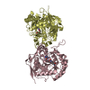
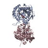
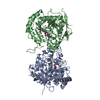
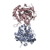
| Title | Catalytic Domain of Human Phenylalanine Hydroxylase Fe(II) in Complex with Tetrahydrobiopterin | ||||||
 Components Components | PHENYLALANINE-4-HYDROXYLASE | ||||||
 Keywords Keywords |  OXIDOREDUCTASE / ferrous iron / 2-His-1-carboxylate facial triad / tetrahydrobiopterin OXIDOREDUCTASE / ferrous iron / 2-His-1-carboxylate facial triad / tetrahydrobiopterin | ||||||
| Function / homology |  Function and homology informationPhenylketonuria / Phenylalanine metabolism / phenylalanine 4-monooxygenase / phenylalanine 4-monooxygenase activity / tyrosine biosynthetic process / catecholamine biosynthetic process / L-phenylalanine catabolic process / amino acid biosynthetic process / iron ion binding / cytosol Function and homology informationPhenylketonuria / Phenylalanine metabolism / phenylalanine 4-monooxygenase / phenylalanine 4-monooxygenase activity / tyrosine biosynthetic process / catecholamine biosynthetic process / L-phenylalanine catabolic process / amino acid biosynthetic process / iron ion binding / cytosolSimilarity search - Function | ||||||
| Biological species |  Homo sapiens (human) Homo sapiens (human) | ||||||
| Method | X-RAY DIFFRACTION / SYNCHROTRON / Resolution: 1.5 Å | ||||||
 Authors Authors | Andersen, O.A. / Flatmark, T. / Hough, E. | ||||||
 Citation Citation | Journal: J.Mol.Biol. / Year: 2001 Title: High resolution crystal structures of the catalytic domain of human phenylalanine hydroxylase in its catalytically active Fe(II) form and binary complex with tetrahydrobiopterin. Authors: Andersen, O.A. / Flatmark, T. / Hough, E. | ||||||
| History |
|
- Structure visualization
| Structure viewer | Molecule: MolmilJmol/JSmol |
|---|
- Downloads & links
-Download
| PDBx/mmCIF format | 1j8u.cif.gz | 148.7 KB | Display | PDBx/mmCIF format |
|---|---|---|---|---|
| PDB format | pdb1j8u.ent.gz | 114.6 KB | Display | PDB format |
| PDBx/mmJSON format | 1j8u.json.gz | Tree view | PDBx/mmJSON format | |
| Others |  Other downloads Other downloads |
-Validation report
| Arichive directory | https://data.pdbj.org/pub/pdb/validation_reports/j8/1j8uftp://data.pdbj.org/pub/pdb/validation_reports/j8/1j8u | HTTPS FTP |
|---|
-Related structure data
| Related structure data |  1j8tC  1pahS C: citing same article ( S: Starting model for refinement |
|---|---|
| Similar structure data |


-Links
 PDBj
PDBj


- Assembly
| Deposited unit | 
| ||||||||
|---|---|---|---|---|---|---|---|---|---|
| 1 | 
| ||||||||
| Unit cell |
|
-Components
| #1: Protein | Mass: 37601.570 Da / Num. of mol.: 1 / Fragment: Catalytic Domain (Residues 103-427) Source method: isolated from a genetically manipulated source Source: (gene. exp.) Homo sapiens (human) / Gene: PAH / Plasmid: pMAL / Production host:  Escherichia coli (E. coli) / References: UniProt: P00439, phenylalanine 4-monooxygenase Escherichia coli (E. coli) / References: UniProt: P00439, phenylalanine 4-monooxygenase |
|---|---|
| #2: Chemical | ChemComp-FE2 /   Mass: 55.845 Da / Num. of mol.: 1 / Source method: obtained synthetically / Formula: Fe Mass: 55.845 Da / Num. of mol.: 1 / Source method: obtained synthetically / Formula: Fe |
| #3: Chemical | ChemComp-H4B / Tetrahydrobiopterin  Mass: 241.247 Da / Num. of mol.: 1 / Source method: obtained synthetically / Formula: C9H15N5O3 / Comment: neurotransmitter*YM Mass: 241.247 Da / Num. of mol.: 1 / Source method: obtained synthetically / Formula: C9H15N5O3 / Comment: neurotransmitter*YM |
| #4: Water | ChemComp-HOH / Water Mass: 18.015 Da / Num. of mol.: 393 / Source method: isolated from a natural source / Formula: H2O Mass: 18.015 Da / Num. of mol.: 393 / Source method: isolated from a natural source / Formula: H2O |
-Experimental details
-Experiment
| Experiment | Method: X-RAY DIFFRACTION / Number of used crystals: 1 |
|---|
- Sample preparation
| Crystal | Density Matthews: 2.98 Å3/Da / Density % sol: 57 % | ||||||||||||||||||||||||||||||||||||||||||
|---|---|---|---|---|---|---|---|---|---|---|---|---|---|---|---|---|---|---|---|---|---|---|---|---|---|---|---|---|---|---|---|---|---|---|---|---|---|---|---|---|---|---|---|
| Crystal grow | Temperature: 277 K / Method: vapor diffusion, hanging drop, anaerobically / pH: 6.8 Details: PEG2000, Na-hepes, ethylene glycol, pH 6.8, Vapor diffusion, hanging drop, anaerobically, temperature 277K | ||||||||||||||||||||||||||||||||||||||||||
| Crystal grow | *PLUS Method: vapor diffusion, hanging drop | ||||||||||||||||||||||||||||||||||||||||||
| Components of the solutions | *PLUS
|
-Data collection
| Diffraction | Mean temperature: 110 K |
|---|---|
| Diffraction source | Source: SYNCHROTRON / Site: ESRF  / Beamline: BM1A / Wavelength: 0.873 Å / Beamline: BM1A / Wavelength: 0.873 Å |
| Detector | Type: MARRESEARCH / Detector: IMAGE PLATE / Date: Sep 30, 2000 |
| Radiation | Protocol: SINGLE WAVELENGTH / Monochromatic (M) / Laue (L): M / Scattering type: x-ray |
| Radiation wavelength | Wavelength: 0.873 Å / Relative weight: 1 |
| Reflection | Resolution: 1.5→20 Å / Num. all: 71940 / Num. obs: 71940 / % possible obs: 99.9 % / Observed criterion σ(F): 0 / Observed criterion σ(I): 0 / Redundancy: 6.9 % / Biso Wilson estimate: 18.4 Å2 / Rmerge(I) obs: 0.051 / Rsym value: 0.051 / Net I/σ(I): 8.7 |
| Reflection shell | Resolution: 1.5→1.58 Å / Redundancy: 5.9 % / Rmerge(I) obs: 0.37 / Mean I/σ(I) obs: 2 / Num. unique all: 10450 / Rsym value: 0.37 / % possible all: 100 |
| Reflection | *PLUS Lowest resolution: 20 Å / Num. measured all: 495560 / Rmerge(I) obs: 0.051 |
| Reflection shell | *PLUS Highest resolution: 1.5 Å / % possible obs: 100 % / Num. possible: 10450 / Num. measured obs: 61233 / Rmerge(I) obs: 0.37 |
- Processing
| Software |
| |||||||||||||||||||||||||
|---|---|---|---|---|---|---|---|---|---|---|---|---|---|---|---|---|---|---|---|---|---|---|---|---|---|---|
| Refinement | Starting model: PDB entry 1PAH Resolution: 1.5→10 Å / Isotropic thermal model: Anisotropic / Cross valid method: THROUGHOUT / σ(F): 0 / σ(I): 0 / Stereochemistry target values: Engh & Huber, 1991
| |||||||||||||||||||||||||
| Displacement parameters | Biso mean: 19.5 Å2 | |||||||||||||||||||||||||
| Refine analyze | Luzzati d res low obs: 5 Å / Luzzati sigma a obs: 0.09 Å | |||||||||||||||||||||||||
| Refinement step | Cycle: LAST / Resolution: 1.5→10 Å
| |||||||||||||||||||||||||
| Refine LS restraints |
| |||||||||||||||||||||||||
| Software | *PLUS Name: SHELXL / Version: 97 / Classification: refinement | |||||||||||||||||||||||||
| Refinement | *PLUS Lowest resolution: 10 Å / Rfactor obs: 0.157 / Rfactor Rfree: 0.203 / Rfactor Rwork: 0.157 | |||||||||||||||||||||||||
| Solvent computation | *PLUS | |||||||||||||||||||||||||
| Displacement parameters | *PLUS | |||||||||||||||||||||||||
| Refine LS restraints | *PLUS
|
