 Movie
Movie Controller


+ Open data

- Basic information
| Entry | Database: PDB / ID: 1j8e | ||||||
|---|---|---|---|---|---|---|---|
| Title | Crystal structure of ligand-binding repeat CR7 from LRP | ||||||
 Components Components | LOW-DENSITY LIPOPROTEIN RECEPTOR-RELATED PROTEIN 1 | ||||||
 Keywords Keywords |  SIGNALING PROTEIN / ligand binding / calcium binding / complement-like repeat / LRP receptor SIGNALING PROTEIN / ligand binding / calcium binding / complement-like repeat / LRP receptor | ||||||
| Function / homology |  Function and homology information Function and homology informationalpha-2 macroglobulin receptor activity / apolipoprotein receptor activity / positive regulation of lipid transport / regulation of phospholipase A2 activity / positive regulation of transcytosis / lipoprotein particle receptor binding / negative regulation of platelet-derived growth factor receptor-beta signaling pathway / negative regulation of metallopeptidase activity / positive regulation of lysosomal protein catabolic process / aorta morphogenesis ...alpha-2 macroglobulin receptor activity / apolipoprotein receptor activity / positive regulation of lipid transport / regulation of phospholipase A2 activity / positive regulation of transcytosis / lipoprotein particle receptor binding / negative regulation of platelet-derived growth factor receptor-beta signaling pathway / negative regulation of metallopeptidase activity / positive regulation of lysosomal protein catabolic process / aorta morphogenesis / negative regulation of smooth muscle cell migration / regulation of cholesterol transport / amyloid-beta clearance by transcytosis / clathrin heavy chain binding / low-density lipoprotein particle receptor activity / regulation of extracellular matrix disassembly / amyloid-beta clearance by cellular catabolic process / scavenger receptor activity / plasma membrane protein complex / positive regulation of amyloid-beta clearance / transcytosis / heparan sulfate proteoglycan binding / astrocyte activation involved in immune response / apoptotic cell clearance / cargo receptor activity / lysosomal transport / retinoid metabolic process / microtubule organizing center / lipoprotein transport / negative regulation of SMAD protein signal transduction / enzyme-linked receptor protein signaling pathway / negative regulation of Wnt signaling pathway / amyloid-beta clearance / positive regulation of endocytosis / apolipoprotein binding / transport across blood-brain barrier / positive regulation of cholesterol efflux / Scavenging of heme from plasma / phagocytosis / clathrin-coated pit / Retinoid metabolism and transport / receptor-mediated endocytosis / regulation of actin cytoskeleton organization / positive regulation of protein localization to plasma membrane / lipid metabolic process / receptor internalization / cellular response to amyloid-beta / endocytic vesicle membrane / positive regulation of protein binding / signaling receptor activity / amyloid-beta binding / basolateral plasma membrane / early endosome / receptor complex / lysosomal membrane / negative regulation of gene expression / focal adhesion / calcium ion binding / protein-containing complex binding / Golgi apparatus / RNA binding / membrane / nucleus / plasma membraneSimilarity search - Function | ||||||
| Biological species |  Homo sapiens (human) Homo sapiens (human) | ||||||
| Method | X-RAY DIFFRACTION / SYNCHROTRON / MOLECULAR REPLACEMENT / Resolution: 1.85 Å | ||||||
 Authors Authors | Simonovic, M. / Dolmer, K. / Huang, W. / Strickland, D.K. / Volz, K. / Gettins, P.G.W. | ||||||
 Citation Citation | Journal: Biochemistry / Year: 2001 Title: Calcium coordination and pH dependence of the calcium affinity of ligand-binding repeat CR7 from the LRP. Comparison with related domains from the LRP and the LDL receptor. Authors: Simonovic, M. / Dolmer, K. / Huang, W. / Strickland, D.K. / Volz, K. / Gettins, P.G. | ||||||
| History |
|
- Structure visualization
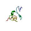
| Structure viewer | Molecule: MolmilJmol/JSmol |
|---|
- Downloads & links
-Download
| PDBx/mmCIF format | 1j8e.cif.gz | 17.6 KB | Display | PDBx/mmCIF format |
|---|---|---|---|---|
| PDB format | pdb1j8e.ent.gz | 12.3 KB | Display | PDB format |
| PDBx/mmJSON format | 1j8e.json.gz | Tree view | PDBx/mmJSON format | |
| Others |  Other downloads Other downloads |
-Validation report
| Arichive directory | https://data.pdbj.org/pub/pdb/validation_reports/j8/1j8eftp://data.pdbj.org/pub/pdb/validation_reports/j8/1j8e | HTTPS FTP |
|---|
-Related structure data


-Links
 PDBj
PDBj


- Assembly
| Deposited unit | 
| ||||||||
|---|---|---|---|---|---|---|---|---|---|
| 1 |
| ||||||||
| Unit cell |
|
-Components
| #1: Protein/peptide | Mass: 4817.019 Da / Num. of mol.: 1 Fragment: COMPLEMENT-LIKE REPEAT 7 (CR7), LDL-RECEPTOR CLASS A 7 Mutation: C1G Source method: isolated from a genetically manipulated source Source: (gene. exp.) Homo sapiens (human) / Plasmid: pGEX-2T / Species (production host): Escherichia coli / Production host:  Escherichia coli BL21(DE3) (bacteria) / Strain (production host): BL21 (DE3) / References: UniProt: Q07954 Escherichia coli BL21(DE3) (bacteria) / Strain (production host): BL21 (DE3) / References: UniProt: Q07954 |
|---|---|
| #2: Chemical | ChemComp-CA /   Mass: 40.078 Da / Num. of mol.: 1 / Source method: obtained synthetically / Formula: Ca Mass: 40.078 Da / Num. of mol.: 1 / Source method: obtained synthetically / Formula: Ca |
| #3: Water | ChemComp-HOH / Water Mass: 18.015 Da / Num. of mol.: 31 / Source method: isolated from a natural source / Formula: H2O Mass: 18.015 Da / Num. of mol.: 31 / Source method: isolated from a natural source / Formula: H2O |
-Experimental details
-Experiment
| Experiment | Method: X-RAY DIFFRACTION / Number of used crystals: 1 |
|---|
- Sample preparation
| Crystal | Density Matthews: 1.84 Å3/Da / Density % sol: 33.03 % | |||||||||||||||||||||||||||||||||||
|---|---|---|---|---|---|---|---|---|---|---|---|---|---|---|---|---|---|---|---|---|---|---|---|---|---|---|---|---|---|---|---|---|---|---|---|---|
| Crystal grow | Temperature: 292 K / Method: vapor diffusion, hanging drop / pH: 3.8 Details: 0.02M Na-acetate, 0.1M CaCl2, 0.3M NaCl, pH 3.8, VAPOR DIFFUSION, HANGING DROP, temperature 292K | |||||||||||||||||||||||||||||||||||
| Crystal grow | *PLUS Temperature: 18 ℃ | |||||||||||||||||||||||||||||||||||
| Components of the solutions | *PLUS
|
-Data collection
| Diffraction | Mean temperature: 200 K |
|---|---|
| Diffraction source | Source: SYNCHROTRON / Site: APS  / Beamline: 14-BM-C / Wavelength: 1 Å / Beamline: 14-BM-C / Wavelength: 1 Å |
| Detector | Type: ADSC QUANTUM 4 / Detector: CCD / Date: Feb 12, 2001 |
| Radiation | Protocol: SINGLE WAVELENGTH / Monochromatic (M) / Laue (L): M / Scattering type: x-ray |
| Radiation wavelength | Wavelength: 1 Å / Relative weight: 1 |
| Reflection | Resolution: 1.85→100 Å / Num. all: 86028 / % possible obs: 88.5 % / Observed criterion σ(I): 0 / Redundancy: 5 % / Biso Wilson estimate: 13.2 Å2 / Rmerge(I) obs: 0.072 / Net I/σ(I): 20 |
| Reflection shell | Resolution: 1.85→1.97 Å / Redundancy: 2 % / Rmerge(I) obs: 0.268 / Num. unique all: 274 / % possible all: 56.6 |
| Reflection | *PLUS Lowest resolution: 100 Å / Num. obs: 2911 / Num. measured all: 86028 |
| Reflection shell | *PLUS % possible obs: 56.6 % / Mean I/σ(I) obs: 4 |
- Processing
| Software |
| ||||||||||||||||||||||||||||||||||||||||
|---|---|---|---|---|---|---|---|---|---|---|---|---|---|---|---|---|---|---|---|---|---|---|---|---|---|---|---|---|---|---|---|---|---|---|---|---|---|---|---|---|---|
| Refinement | Method to determine structure: MOLECULAR REPLACEMENT Starting model: Composite search model constructed based on coordinates of 1AJJ, 1D2L, and 1CR8. Resolution: 1.85→21.96 Å / Rfactor Rfree error: 0.013 / Data cutoff high absF: 1341106.64 / Data cutoff low absF: 0 / Isotropic thermal model: RESTRAINED / Cross valid method: THROUGHOUT / σ(F): 0 / Stereochemistry target values: Engh & Huber
| ||||||||||||||||||||||||||||||||||||||||
| Solvent computation | Solvent model: FLAT MODEL / Bsol: 51.37 Å2 / ksol: 0.419 e/Å3 | ||||||||||||||||||||||||||||||||||||||||
| Displacement parameters | Biso mean: 21 Å2
| ||||||||||||||||||||||||||||||||||||||||
| Refine analyze |
| ||||||||||||||||||||||||||||||||||||||||
| Refinement step | Cycle: LAST / Resolution: 1.85→21.96 Å
| ||||||||||||||||||||||||||||||||||||||||
| Refine LS restraints |
| ||||||||||||||||||||||||||||||||||||||||
| LS refinement shell | Resolution: 1.85→1.97 Å / Rfactor Rfree error: 0.054 / Total num. of bins used: 6
| ||||||||||||||||||||||||||||||||||||||||
| Xplor file |
| ||||||||||||||||||||||||||||||||||||||||
| Software | *PLUS Name: CNS / Version: 1 / Classification: refinement | ||||||||||||||||||||||||||||||||||||||||
| Refinement | *PLUS Highest resolution: 1.85 Å / Lowest resolution: 21.96 Å / σ(F): 0 / % reflection Rfree: 9.7 % / Rfactor obs: 0.186 | ||||||||||||||||||||||||||||||||||||||||
| Solvent computation | *PLUS | ||||||||||||||||||||||||||||||||||||||||
| Displacement parameters | *PLUS Biso mean: 21 Å2 | ||||||||||||||||||||||||||||||||||||||||
| Refine LS restraints | *PLUS
| ||||||||||||||||||||||||||||||||||||||||
| LS refinement shell | *PLUS Rfactor Rfree: 0.288 / % reflection Rfree: 9.3 % / Rfactor Rwork: 0.227 |
