 Movie
Movie Controller


+ Open data

- Basic information
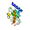
| Entry | Database: PDB / ID: 1j7d | ||||||
|---|---|---|---|---|---|---|---|
| Title | Crystal Structure of hMms2-hUbc13 | ||||||
 Components Components |
| ||||||
 Keywords Keywords | UNKNOWN FUNCTION /  Ubiquitin / Ubc / DNA repair / Traf6 / NFkB Ubiquitin / Ubc / DNA repair / Traf6 / NFkB | ||||||
| Function / homology |  Function and homology information Function and homology informationerror-free postreplication DNA repair / : / UBC13-MMS2 complex / ubiquitin conjugating enzyme complex / ubiquitin-protein transferase activator activity / positive regulation of protein K63-linked ubiquitination / DNA double-strand break processing / postreplication repair / positive regulation of double-strand break repair / positive regulation of intracellular signal transduction ...error-free postreplication DNA repair / : / UBC13-MMS2 complex / ubiquitin conjugating enzyme complex / ubiquitin-protein transferase activator activity / positive regulation of protein K63-linked ubiquitination / DNA double-strand break processing / postreplication repair / positive regulation of double-strand break repair / positive regulation of intracellular signal transduction / E2 ubiquitin-conjugating enzyme / ubiquitin conjugating enzyme activity / protein K63-linked ubiquitination / antiviral innate immune response / regulation of DNA repair / ubiquitin ligase complex / negative regulation of TORC1 signaling / IRAK1 recruits IKK complex / IRAK1 recruits IKK complex upon TLR7/8 or 9 stimulation / positive regulation of DNA repair / TRAF6 mediated IRF7 activation in TLR7/8 or 9 signaling / TICAM1, RIP1-mediated IKK complex recruitment / IKK complex recruitment mediated by RIP1 / JNK (c-Jun kinases) phosphorylation and activation mediated by activated human TAK1 / ubiquitin binding / activated TAK1 mediates p38 MAPK activation / Nonhomologous End-Joining (NHEJ) / double-strand break repair via homologous recombination / TAK1-dependent IKK and NF-kappa-B activation / NOD1/2 Signaling Pathway / G2/M DNA damage checkpoint / ISG15 antiviral mechanism / CLEC7A (Dectin-1) signaling / Formation of Incision Complex in GG-NER / FCERI mediated NF-kB activation / Aggrephagy / Interleukin-1 signaling / protein polyubiquitination / ubiquitin-protein transferase activity / Antigen processing: Ubiquitination & Proteasome degradation / Downstream TCR signaling / E3 ubiquitin ligases ubiquitinate target proteins / Recruitment and ATM-mediated phosphorylation of repair and signaling proteins at DNA double strand breaks / positive regulation of NF-kappaB transcription factor activity / T cell receptor signaling pathway / Processing of DNA double-strand break ends / proteasome-mediated ubiquitin-dependent protein catabolic process / positive regulation of canonical NF-kappaB signal transduction / protein ubiquitination / ubiquitin protein ligase binding / SARS-CoV-2 activates/modulates innate and adaptive immune responses / protein-containing complex / RNA binding / extracellular exosome / nucleoplasm / ATP binding / nucleus / cytosol / cytoplasmSimilarity search - Function | ||||||
| Biological species |  Homo sapiens (human) Homo sapiens (human) | ||||||
| Method | X-RAY DIFFRACTION / SYNCHROTRON / MAD / Resolution: 1.85 Å | ||||||
 Authors Authors | Moraes, T.F. / Edwards, R.A. / McKenna, S. / Pashushok, L. / Xiao, W. / Glover, J.N.M. / Ellison, M.J. | ||||||
 Citation Citation | Journal: Nat.Struct.Biol. / Year: 2001 Title: Crystal structure of the human ubiquitin conjugating enzyme complex, hMms2-hUbc13. Authors: Moraes, T.F. / Edwards, R.A. / McKenna, S. / Pastushok, L. / Xiao, W. / Glover, J.N. / Ellison, M.J. | ||||||
| History |
|
- Structure visualization
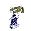
| Structure viewer | Molecule: MolmilJmol/JSmol |
|---|
- Downloads & links
-Download
| PDBx/mmCIF format | 1j7d.cif.gz | 71.9 KB | Display | PDBx/mmCIF format |
|---|---|---|---|---|
| PDB format | pdb1j7d.ent.gz | 54 KB | Display | PDB format |
| PDBx/mmJSON format | 1j7d.json.gz | Tree view | PDBx/mmJSON format | |
| Others |  Other downloads Other downloads |
-Validation report
| Arichive directory | https://data.pdbj.org/pub/pdb/validation_reports/j7/1j7dftp://data.pdbj.org/pub/pdb/validation_reports/j7/1j7d | HTTPS FTP |
|---|
-Related structure data


-Links
 PDBj
PDBj


- Assembly
| Deposited unit | 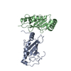
| ||||||||
|---|---|---|---|---|---|---|---|---|---|
| 1 |
| ||||||||
| Unit cell |
|
-Components
| #1: Protein | Mass: 16380.731 Da / Num. of mol.: 1 Source method: isolated from a genetically manipulated source Source: (gene. exp.) Homo sapiens (human) / Production host:  Escherichia coli (E. coli) / References: UniProt: Q15819 Escherichia coli (E. coli) / References: UniProt: Q15819 |
|---|---|
| #2: Protein | Mass: 17157.770 Da / Num. of mol.: 1 Source method: isolated from a genetically manipulated source Source: (gene. exp.) Homo sapiens (human) / Production host: Escherichia coli (E. coli) / References: UniProt: P61088, ubiquitin-protein ligase |
| #3: Water | ChemComp-HOH / Water Mass: 18.015 Da / Num. of mol.: 190 / Source method: isolated from a natural source / Formula: H2O Mass: 18.015 Da / Num. of mol.: 190 / Source method: isolated from a natural source / Formula: H2O |
-Experimental details
-Experiment
| Experiment | Method: X-RAY DIFFRACTION / Number of used crystals: 1 |
|---|
- Sample preparation
| Crystal | Density Matthews: 2.23 Å3/Da / Density % sol: 44.83 % | ||||||||||||||||||||||||||||||||||||||||||||||||
|---|---|---|---|---|---|---|---|---|---|---|---|---|---|---|---|---|---|---|---|---|---|---|---|---|---|---|---|---|---|---|---|---|---|---|---|---|---|---|---|---|---|---|---|---|---|---|---|---|---|
| Crystal grow | Temperature: 298 K / Method: vapor diffusion, hanging drop / pH: 6.5 Details: 20% PEG 8000, 100mM Citrate, pH 6.5, VAPOR DIFFUSION, HANGING DROP, temperature 298K | ||||||||||||||||||||||||||||||||||||||||||||||||
| Crystal grow | *PLUS Temperature: 20-24 ℃ / pH: 7.5 | ||||||||||||||||||||||||||||||||||||||||||||||||
| Components of the solutions | *PLUS
|
-Data collection
| Diffraction | Mean temperature: 100 K | ||||||||||||
|---|---|---|---|---|---|---|---|---|---|---|---|---|---|
| Diffraction source | Source: SYNCHROTRON / Site: NSLS  / Beamline: X12C / Wavelength: 0.981958, 0.982127, 1.022130 / Beamline: X12C / Wavelength: 0.981958, 0.982127, 1.022130 | ||||||||||||
| Detector | Type: BRANDEIS - B1 / Detector: CCD / Date: Oct 10, 2000 | ||||||||||||
| Radiation | Protocol: SINGLE WAVELENGTH / Monochromatic (M) / Laue (L): M / Scattering type: x-ray | ||||||||||||
| Radiation wavelength |
| ||||||||||||
| Reflection | Resolution: 1.85→40 Å / Num. all: 26066 / Num. obs: 26066 / % possible obs: 98.3 % / Redundancy: 4.25 % / Biso Wilson estimate: 23.4 Å2 / Rmerge(I) obs: 0.034 / Rsym value: 0.034 / Net I/σ(I): 37.1 | ||||||||||||
| Reflection shell | Resolution: 1.85→1.88 Å / Redundancy: 2.5 % / Rmerge(I) obs: 0.263 / Mean I/σ(I) obs: 3.2 / Num. unique all: 1161 / Rsym value: 0.263 / % possible all: 89.5 | ||||||||||||
| Reflection | *PLUS Lowest resolution: 40 Å / Num. measured all: 110669 / Rmerge(I) obs: 0.024 |
- Processing
| Software |
| |||||||||||||||||||||||||
|---|---|---|---|---|---|---|---|---|---|---|---|---|---|---|---|---|---|---|---|---|---|---|---|---|---|---|
| Refinement | Method to determine structure: MAD / Resolution: 1.85→40 Å / Stereochemistry target values: CNS targets
| |||||||||||||||||||||||||
| Refinement step | Cycle: LAST / Resolution: 1.85→40 Å
| |||||||||||||||||||||||||
| Refine LS restraints |
| |||||||||||||||||||||||||
| LS refinement shell | Resolution: 1.85→1.87 Å
| |||||||||||||||||||||||||
| Software | *PLUS Name: CNS / Classification: refinement | |||||||||||||||||||||||||
| Refinement | *PLUS Lowest resolution: 40 Å / % reflection Rfree: 5 % / Rfactor obs: 0.215 | |||||||||||||||||||||||||
| Solvent computation | *PLUS | |||||||||||||||||||||||||
| Displacement parameters | *PLUS |
