 Movie
Movie Controller


+ Open data

- Basic information
| Entry | Database: PDB / ID: 1j58 | ||||||
|---|---|---|---|---|---|---|---|
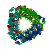
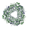
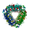
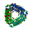
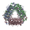
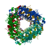
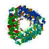
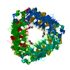
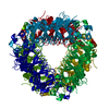
| Title | Crystal Structure of Oxalate Decarboxylase | ||||||
 Components Components | YVRK PROTEIN | ||||||
 Keywords Keywords | METAL BINDING PROTEIN /  CUPIN / DECARBOXYKLASE / OXALATE / MANGANESE / FORMATE CUPIN / DECARBOXYKLASE / OXALATE / MANGANESE / FORMATE | ||||||
| Function / homology |  Function and homology informationoxalate decarboxylase / oxalate decarboxylase activity / oxalate metabolic process / metal ion binding / cytoplasm Function and homology informationoxalate decarboxylase / oxalate decarboxylase activity / oxalate metabolic process / metal ion binding / cytoplasmSimilarity search - Function | ||||||
| Biological species |  Bacillus subtilis (bacteria) Bacillus subtilis (bacteria) | ||||||
| Method | X-RAY DIFFRACTION / SYNCHROTRON / Resolution: 1.75 Å | ||||||
 Authors Authors | Anand, R. / Dorrestein, P.C. / Kinsland, C. / Begley, T.P. / Ealick, S.E. | ||||||
 Citation Citation | Journal: Biochemistry / Year: 2002 Title: Structure of oxalate decarboxylase from Bacillus subtilis at 1.75 A resolution. Authors: Anand, R. / Dorrestein, P.C. / Kinsland, C. / Begley, T.P. / Ealick, S.E. | ||||||
| History |
|
- Structure visualization
| Structure viewer | Molecule: MolmilJmol/JSmol |
|---|
- Downloads & links
-Download
| PDBx/mmCIF format | 1j58.cif.gz | 98.2 KB | Display | PDBx/mmCIF format |
|---|---|---|---|---|
| PDB format | pdb1j58.ent.gz | 73.8 KB | Display | PDB format |
| PDBx/mmJSON format | 1j58.json.gz | Tree view | PDBx/mmJSON format | |
| Others |  Other downloads Other downloads |
-Validation report
| Arichive directory | https://data.pdbj.org/pub/pdb/validation_reports/j5/1j58ftp://data.pdbj.org/pub/pdb/validation_reports/j5/1j58 | HTTPS FTP |
|---|
-Related structure data


-Links
 PDBj
PDBj- Assembly
| Deposited unit | 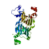
| ||||||||
|---|---|---|---|---|---|---|---|---|---|
| 1 | x 6
| ||||||||
| Unit cell |
| ||||||||
| Details | The biological assembly is a hexamer constructed by doing a three fold and a two fold symmetry operation on chain A |
-Components
| #1: Protein | Mass: 44042.957 Da / Num. of mol.: 1 Source method: isolated from a genetically manipulated source Source: (gene. exp.) Bacillus subtilis (bacteria) / Plasmid: PET16b / Production host: Bacteria (eubacteria) / References: UniProt: O34714 | ||||||
|---|---|---|---|---|---|---|---|
| #2: Chemical |   Mass: 54.938 Da / Num. of mol.: 2 / Source method: obtained synthetically / Formula: Mn Mass: 54.938 Da / Num. of mol.: 2 / Source method: obtained synthetically / Formula: Mn#3: Chemical | ChemComp-MG / |   Mass: 24.305 Da / Num. of mol.: 1 / Source method: obtained synthetically / Formula: Mg Mass: 24.305 Da / Num. of mol.: 1 / Source method: obtained synthetically / Formula: Mg#4: Chemical | ChemComp-FMT / | Formic acid  Mass: 46.025 Da / Num. of mol.: 1 / Source method: obtained synthetically / Formula: CH2O2 Mass: 46.025 Da / Num. of mol.: 1 / Source method: obtained synthetically / Formula: CH2O2#5: Water | ChemComp-HOH / | Water Mass: 18.015 Da / Num. of mol.: 439 / Source method: isolated from a natural source / Formula: H2O Mass: 18.015 Da / Num. of mol.: 439 / Source method: isolated from a natural source / Formula: H2O |
-Experimental details
-Experiment
| Experiment | Method: X-RAY DIFFRACTION / Number of used crystals: 1 |
|---|
- Sample preparation
| Crystal | Density Matthews: 3.23 Å3/Da / Density % sol: 61.95 % | ||||||||||||||||||||||||
|---|---|---|---|---|---|---|---|---|---|---|---|---|---|---|---|---|---|---|---|---|---|---|---|---|---|
| Crystal grow | Temperature: 298 K / Method: vapor diffusion / pH: 8 Details: PEG 4000, Tris-Hcl, sodium chloride, pH 8.0, VAPOR DIFFUSION, temperature 298K | ||||||||||||||||||||||||
| Crystal | *PLUS Density % sol: 60 % | ||||||||||||||||||||||||
| Crystal grow | *PLUS Method: vapor diffusion, hanging drop | ||||||||||||||||||||||||
| Components of the solutions | *PLUS
|
-Data collection
| Diffraction |
| |||||||||||||||
|---|---|---|---|---|---|---|---|---|---|---|---|---|---|---|---|---|
| Diffraction source | Source: SYNCHROTRON / Site: CHESS  / Beamline: F2 / Wavelength: .9791, 0.9794, 0.9641, 0.9807 / Beamline: F2 / Wavelength: .9791, 0.9794, 0.9641, 0.9807 | |||||||||||||||
| Detector | Type: ADSC QUANTUM 4 / Detector: CCD / Date: May 15, 2001 | |||||||||||||||
| Radiation | Protocol: SINGLE WAVELENGTH / Monochromatic (M) / Laue (L): M / Scattering type: x-ray | |||||||||||||||
| Radiation wavelength |
| |||||||||||||||
| Reflection | Resolution: 1.75→29.83 Å / Num. all: 57048 / Num. obs: 57048 / % possible obs: 100 % / Observed criterion σ(F): 0 / Observed criterion σ(I): 0 / Redundancy: 4.4 % / Biso Wilson estimate: 11.7 Å2 / Rmerge(I) obs: 0.083 / Net I/σ(I): 7.1 | |||||||||||||||
| Reflection shell | Resolution: 1.75→1.85 Å / Redundancy: 3.1 % / Rmerge(I) obs: 0.23 / % possible all: 99.7 | |||||||||||||||
| Reflection | *PLUS Num. obs: 57307 / % possible obs: 99.9 % / Redundancy: 7.1 % / Num. measured all: 406760 / Rmerge(I) obs: 0.083 | |||||||||||||||
| Reflection shell | *PLUS Lowest resolution: 1.84 Å / % possible obs: 99.7 % / Rmerge(I) obs: 0.325 / Mean I/σ(I) obs: 2.3 |
- Processing
| Software |
| ||||||||||||||||||||||||||||||||||||
|---|---|---|---|---|---|---|---|---|---|---|---|---|---|---|---|---|---|---|---|---|---|---|---|---|---|---|---|---|---|---|---|---|---|---|---|---|---|
| Refinement | Resolution: 1.75→29.83 Å / Rfactor Rfree error: 0.003 / Data cutoff high absF: 3802941.91 / Data cutoff high rms absF: 3802941.91 / Data cutoff low absF: 0 / Isotropic thermal model: RESTRAINED / Cross valid method: THROUGHOUT / σ(F): 0 / σ(I): 0 / Stereochemistry target values: Engh & Huber
| ||||||||||||||||||||||||||||||||||||
| Solvent computation | Solvent model: FLAT MODEL / Bsol: 55.7172 Å2 / ksol: 0.387726 e/Å3 | ||||||||||||||||||||||||||||||||||||
| Displacement parameters | Biso mean: 15.3 Å2
| ||||||||||||||||||||||||||||||||||||
| Refine analyze |
| ||||||||||||||||||||||||||||||||||||
| Refinement step | Cycle: LAST / Resolution: 1.75→29.83 Å
| ||||||||||||||||||||||||||||||||||||
| Refine LS restraints |
| ||||||||||||||||||||||||||||||||||||
| LS refinement shell | Resolution: 1.75→1.86 Å / Rfactor Rfree error: 0.009 / Total num. of bins used: 6
| ||||||||||||||||||||||||||||||||||||
| Xplor file |
| ||||||||||||||||||||||||||||||||||||
| Refinement | *PLUS Lowest resolution: 25 Å / Num. reflection obs: 57039 / % reflection Rfree: 10 % / Rfactor all: 0.192 / Rfactor obs: 0.181 / Rfactor Rfree: 0.1784 / Rfactor Rwork: 0.1831 | ||||||||||||||||||||||||||||||||||||
| Solvent computation | *PLUS | ||||||||||||||||||||||||||||||||||||
| Displacement parameters | *PLUS | ||||||||||||||||||||||||||||||||||||
| Refine LS restraints | *PLUS
| ||||||||||||||||||||||||||||||||||||
| LS refinement shell | *PLUS Rfactor Rfree: 0.241 / Rfactor Rwork: 0.228 |
