 Movie
Movie Controller

[English] 日本語
 Yorodumi
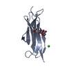
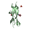
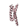
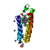
- PDB-1j27: Crystal structure of a hypothetical protein, TT1725, from Thermus... -
+ Open data

- Basic information
| Entry | Database: PDB / ID: 1j27 | ||||||
|---|---|---|---|---|---|---|---|
| Title | Crystal structure of a hypothetical protein, TT1725, from Thermus thermophilus HB8 at 1.7A resolution | ||||||
 Components Components | hypothetical protein TT1725 Hypothesis Hypothesis | ||||||
 Keywords Keywords | structural genomics / unknown function / hypothetical protein from Thermus thermophilus HB8 / MAD / RIKEN Structural Genomics/Proteomics Initiative / RSGI | ||||||
| Function / homology | TT1725-like / Protein of unknown function DUF503 / TT1725-like superfamily / Protein of unknown function (DUF503) / Alpha-Beta Plaits / 2-Layer Sandwich / Alpha Beta / Uncharacterized protein Function and homology information Function and homology information | ||||||
| Biological species |   Thermus thermophilus (bacteria) Thermus thermophilus (bacteria) | ||||||
| Method | X-RAY DIFFRACTION / SYNCHROTRON / MAD / Resolution: 1.7 Å | ||||||
 Authors Authors | Seto, A. / Shirouzu, M. / Terada, T. / Murayama, K. / Kuramitsu, S. / Yokoyama, S. / RIKEN Structural Genomics/Proteomics Initiative (RSGI) | ||||||
 Citation Citation | Journal: Proteins / Year: 2003 Title: Crystal structure of a hypothetical protein, TT1725, from Thermus thermophilus HB8 at 1.7 A resolution Authors: Seto, A. / Shirouzu, M. / Terada, T. / Murayama, K. / Kuramitsu, S. / Yokoyama, S. | ||||||
| History |
|
- Structure visualization
| Structure viewer | Molecule: MolmilJmol/JSmol |
|---|
- Downloads & links
-Download
| PDBx/mmCIF format | 1j27.cif.gz | 33.5 KB | Display | PDBx/mmCIF format |
|---|---|---|---|---|
| PDB format | pdb1j27.ent.gz | 22.8 KB | Display | PDB format |
| PDBx/mmJSON format | 1j27.json.gz | Tree view | PDBx/mmJSON format | |
| Others |  Other downloads Other downloads |
-Validation report
| Arichive directory | https://data.pdbj.org/pub/pdb/validation_reports/j2/1j27ftp://data.pdbj.org/pub/pdb/validation_reports/j2/1j27 | HTTPS FTP |
|---|
-Related structure data
| Similar structure data | |
|---|---|
| Other databases |


-Links
 PDBj
PDBj- Assembly
| Deposited unit | 
| ||||||||
|---|---|---|---|---|---|---|---|---|---|
| 1 |
| ||||||||
| 2 | 
| ||||||||
| Unit cell |
|
-Components
| #1: Protein | Hypothesis Mass: 11421.213 Da / Num. of mol.: 1 Source method: isolated from a genetically manipulated source Source: (gene. exp.) Thermus thermophilus (bacteria) / Strain: HB8 / Gene: TT1725 / Plasmid: pET3a / Production host: Escherichia coli (E. coli) / References: UniProt: Q84BR1 |
|---|---|
| #2: Water | ChemComp-HOH / Water Mass: 18.015 Da / Num. of mol.: 129 / Source method: isolated from a natural source / Formula: H2O Mass: 18.015 Da / Num. of mol.: 129 / Source method: isolated from a natural source / Formula: H2O |
-Experimental details
-Experiment
| Experiment | Method: X-RAY DIFFRACTION |
|---|
- Sample preparation
| Crystal | Density Matthews: 2.75 Å3/Da / Density % sol: 55.33 % | ||||||||||||||||||||||||||||||
|---|---|---|---|---|---|---|---|---|---|---|---|---|---|---|---|---|---|---|---|---|---|---|---|---|---|---|---|---|---|---|---|
| Crystal grow | *PLUS Temperature: 20 ℃ / pH: 8.5 / Method: vapor diffusion, hanging drop | ||||||||||||||||||||||||||||||
| Components of the solutions | *PLUS
|
-Data collection
| Diffraction source | Source: SYNCHROTRON / Site: SPring-8  / Beamline: BL44B2 / Beamline: BL44B2 |
|---|---|
| Radiation | Protocol: MAD / Monochromatic (M) / Laue (L): M / Scattering type: x-ray |
| Radiation wavelength | Relative weight: 1 |
| Reflection | Resolution: 1.7→50 Å / Num. obs: 15362 / Observed criterion σ(I): 0 / Biso Wilson estimate: 11.7 Å2 |
| Reflection | *PLUS Highest resolution: 1.7 Å / % possible obs: 98.7 % / Num. measured all: 71421 / Rmerge(I) obs: 0.028 |
| Reflection shell | *PLUS Highest resolution: 1.7 Å / Lowest resolution: 1.76 Å / % possible obs: 89.6 % / Rmerge(I) obs: 0.097 / Mean I/σ(I) obs: 13.8 |
- Processing
| Software | Name: CNS / Version: 1.1 / Classification: refinement | |||||||||||||||||||||||||
|---|---|---|---|---|---|---|---|---|---|---|---|---|---|---|---|---|---|---|---|---|---|---|---|---|---|---|
| Refinement | Method to determine structure: MAD / Resolution: 1.7→21.48 Å / Rfactor Rfree error: 0.006 / Data cutoff high absF: 1149962.57 / Data cutoff low absF: 0 / Isotropic thermal model: RESTRAINED / Cross valid method: THROUGHOUT / σ(F): 0
| |||||||||||||||||||||||||
| Solvent computation | Solvent model: FLAT MODEL / Bsol: 65.5761 Å2 / ksol: 0.451101 e/Å3 | |||||||||||||||||||||||||
| Displacement parameters | Biso mean: 14.2 Å2
| |||||||||||||||||||||||||
| Refine analyze |
| |||||||||||||||||||||||||
| Refinement step | Cycle: LAST / Resolution: 1.7→21.48 Å
| |||||||||||||||||||||||||
| Refine LS restraints |
| |||||||||||||||||||||||||
| LS refinement shell | Resolution: 1.7→1.81 Å / Rfactor Rfree error: 0.014 / Total num. of bins used: 6
| |||||||||||||||||||||||||
| Xplor file |
| |||||||||||||||||||||||||
| Refinement | *PLUS Highest resolution: 1.7 Å / % reflection Rfree: 10 % | |||||||||||||||||||||||||
| Solvent computation | *PLUS | |||||||||||||||||||||||||
| Displacement parameters | *PLUS | |||||||||||||||||||||||||
| Refine LS restraints | *PLUS
|
