 Movie
Movie Controller


+ Open data

- Basic information
| Entry | Database: PDB / ID: 1iw9 | |||||||||
|---|---|---|---|---|---|---|---|---|---|---|
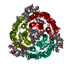
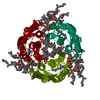
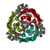
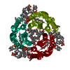
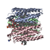
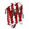
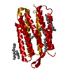
| Title | Crystal Structure of the M Intermediate of Bacteriorhodopsin | |||||||||
 Components Components | bacteriorhodopsin | |||||||||
 Keywords Keywords | PROTON TRANSPORT / 7 Transmembrane Helices / RIKEN Structural Genomics/Proteomics Initiative / RSGI / Structural Genomics | |||||||||
| Function / homology |  Function and homology informationphotoreceptor activity / phototransduction / proton transmembrane transport / monoatomic ion channel activity / plasma membrane Function and homology informationphotoreceptor activity / phototransduction / proton transmembrane transport / monoatomic ion channel activity / plasma membraneSimilarity search - Function | |||||||||
| Biological species |  Halobacterium salinarum (Halophile) Halobacterium salinarum (Halophile) | |||||||||
| Method | X-RAY DIFFRACTION / SYNCHROTRON / MOLECULAR REPLACEMENT / Resolution: 2.5 Å | |||||||||
 Authors Authors | Takeda, K. / Matsui, Y. / Kamiya, N. / Adachi, S. / Okumura, H. / Kouyama, T. / RIKEN Structural Genomics/Proteomics Initiative (RSGI) | |||||||||
 Citation Citation | Journal: J.Mol.Biol. / Year: 2004 Title: Crystal structure of the M intermediate of bacteriorhodopsin: allosteric structural changes mediated by sliding movement of a transmembrane helix Authors: Takeda, K. / Matsui, Y. / Kamiya, N. / Adachi, S. / Okumura, H. / Kouyama, T. #1: Journal: J.Mol.Biol. / Year: 2004Title: Crystal Structure of the L Intermediate of Bacteriorhodopsin: Evidence for Vertical Translocation of a Water Molecule during the Proton Pumping Cycle Authors: Kouyama, T. / Nishikawa, T. / Tokuhisa, T. / Okumura, H. | |||||||||
| History |
|
- Structure visualization
| Structure viewer | Molecule: MolmilJmol/JSmol |
|---|
- Downloads & links
-Download
| PDBx/mmCIF format | 1iw9.cif.gz | 68.5 KB | Display | PDBx/mmCIF format |
|---|---|---|---|---|
| PDB format | pdb1iw9.ent.gz | 48.4 KB | Display | PDB format |
| PDBx/mmJSON format | 1iw9.json.gz | Tree view | PDBx/mmJSON format | |
| Others |  Other downloads Other downloads |
-Validation report
| Arichive directory | https://data.pdbj.org/pub/pdb/validation_reports/iw/1iw9ftp://data.pdbj.org/pub/pdb/validation_reports/iw/1iw9 | HTTPS FTP |
|---|
-Related structure data
| Related structure data | 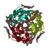 1dzeSC S: Starting model for refinement C: citing same article ( |
|---|---|
| Similar structure data | |
| Other databases |


-Links
 PDBj
PDBj


- Assembly
| Deposited unit | 
| ||||||||
|---|---|---|---|---|---|---|---|---|---|
| 1 | 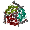
| ||||||||
| Unit cell |
| ||||||||
| Details | The biological assembly is a trimer generated from the monomer in the asymmetric unit by the operations:1-y, x-y, z and 1-x+y, 1-x, z |
-Components
-Protein / Sugars , 2 types, 2 molecules A
| #1: Protein | Mass: 26814.412 Da / Num. of mol.: 1 / Source method: isolated from a natural source / Source: (natural) Halobacterium salinarum (Halophile) / Strain: jw3 / References: UniProt: P02945 |
|---|---|
| #2: Polysaccharide | beta-D-galactopyranose-(1-6)-alpha-D-mannopyranose-(1-2)-alpha-D-glucopyranose / Mass: 504.438 Da / Num. of mol.: 1 Source method: isolated from a genetically manipulated source |
-Non-polymers , 4 types, 31 molecules 

| #3: Chemical | ChemComp-RET / Retinal Mass: 284.436 Da / Num. of mol.: 1 / Source method: obtained synthetically / Formula: C20H28O Mass: 284.436 Da / Num. of mol.: 1 / Source method: obtained synthetically / Formula: C20H28O | ||||
|---|---|---|---|---|---|
| #4: Chemical | ChemComp-L3P /  Mass: 885.179 Da / Num. of mol.: 4 / Source method: obtained synthetically / Formula: C46H94O11P2 Mass: 885.179 Da / Num. of mol.: 4 / Source method: obtained synthetically / Formula: C46H94O11P2#5: Chemical | ChemComp-L2P / |  Mass: 653.157 Da / Num. of mol.: 1 / Source method: obtained synthetically / Formula: C43H88O3 Mass: 653.157 Da / Num. of mol.: 1 / Source method: obtained synthetically / Formula: C43H88O3#6: Water | ChemComp-HOH / | WaterMass: 18.015 Da / Num. of mol.: 25 / Source method: isolated from a natural source / Formula: H2O |
-Experimental details
-Experiment
| Experiment | Method: X-RAY DIFFRACTION / Number of used crystals: 3 |
|---|
- Sample preparation
| Crystal | Density Matthews: 3.16 Å3/Da / Density % sol: 61.11 % | |||||||||||||||||||||||||||||||||||||||||||||||||||||||||||||||
|---|---|---|---|---|---|---|---|---|---|---|---|---|---|---|---|---|---|---|---|---|---|---|---|---|---|---|---|---|---|---|---|---|---|---|---|---|---|---|---|---|---|---|---|---|---|---|---|---|---|---|---|---|---|---|---|---|---|---|---|---|---|---|---|---|
| Crystal grow | Temperature: 293 K / Method: vapor diffusion, sitting drop / pH: 5.2 Details: ammonium sulfate, pH 5.2, VAPOR DIFFUSION, SITTING DROP, temperature 293K | |||||||||||||||||||||||||||||||||||||||||||||||||||||||||||||||
| Crystal grow | *PLUS Temperature: 10 ℃ / Method: vapor diffusion, sitting drop | |||||||||||||||||||||||||||||||||||||||||||||||||||||||||||||||
| Components of the solutions | *PLUS
|
-Data collection
| Diffraction | Mean temperature: 100 K |
|---|---|
| Diffraction source | Source: SYNCHROTRON / Site: SPring-8  / Beamline: BL44B2 / Wavelength: 0.708 Å / Beamline: BL44B2 / Wavelength: 0.708 Å |
| Detector | Type: RIGAKU / Detector: IMAGE PLATE / Date: Nov 18, 1998 / Details: mirrors |
| Radiation | Monochromator: DOUBLE-CRYSTAL / Protocol: SINGLE WAVELENGTH / Monochromatic (M) / Laue (L): M / Scattering type: x-ray |
| Radiation wavelength | Wavelength: 0.708 Å / Relative weight: 1 |
| Reflection | Resolution: 2.5→50 Å / Num. all: 12434 / Num. obs: 11480 / % possible obs: 91.7 % / Observed criterion σ(F): 0 / Observed criterion σ(I): 0 / Redundancy: 14.4 % / Rmerge(I) obs: 0.069 |
| Reflection shell | Resolution: 2.5→2.59 Å / Rsym value: 0.304 / % possible all: 55.2 |
| Reflection | *PLUS Lowest resolution: 50 Å / Num. obs: 10066 / % possible obs: 92.1 % / Redundancy: 5.9 % / Rmerge(I) obs: 0.068 |
| Reflection shell | *PLUS % possible obs: 92.1 % / Rmerge(I) obs: 0.522 / Mean I/σ(I) obs: 1.4 |
- Processing
| Software |
| |||||||||||||||||||||||||||||||||||||||||||||||||
|---|---|---|---|---|---|---|---|---|---|---|---|---|---|---|---|---|---|---|---|---|---|---|---|---|---|---|---|---|---|---|---|---|---|---|---|---|---|---|---|---|---|---|---|---|---|---|---|---|---|---|
| Refinement | Method to determine structure: MOLECULAR REPLACEMENT Starting model: PDB ENTRY 1dze Resolution: 2.5→15.3 Å / Isotropic thermal model: Overall / Cross valid method: troughout / σ(F): 0 / σ(I): 0 / Stereochemistry target values: Engh & Huber
| |||||||||||||||||||||||||||||||||||||||||||||||||
| Displacement parameters | Biso mean: 47 Å2
| |||||||||||||||||||||||||||||||||||||||||||||||||
| Refine analyze |
| |||||||||||||||||||||||||||||||||||||||||||||||||
| Refinement step | Cycle: LAST / Resolution: 2.5→15.3 Å
| |||||||||||||||||||||||||||||||||||||||||||||||||
| Refine LS restraints |
| |||||||||||||||||||||||||||||||||||||||||||||||||
| LS refinement shell |
| |||||||||||||||||||||||||||||||||||||||||||||||||
| Refinement | *PLUS Lowest resolution: 28 Å / % reflection Rfree: 5 % / Rfactor Rfree: 0.286 / Rfactor Rwork: 0.236 | |||||||||||||||||||||||||||||||||||||||||||||||||
| Solvent computation | *PLUS | |||||||||||||||||||||||||||||||||||||||||||||||||
| Displacement parameters | *PLUS | |||||||||||||||||||||||||||||||||||||||||||||||||
| Refine LS restraints | *PLUS
|
