 Movie
Movie Controller

[English] 日本語
 Yorodumi
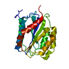
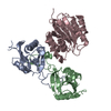
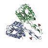
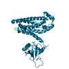
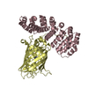
- PDB-1ijk: The von Willebrand Factor mutant (I546V) A1 domain-botrocetin Complex -
+ Open data

- Basic information
| Entry | Database: PDB / ID: 1ijk | ||||||
|---|---|---|---|---|---|---|---|
| Title | The von Willebrand Factor mutant (I546V) A1 domain-botrocetin Complex | ||||||
 Components Components |
| ||||||
 Keywords Keywords | BLOOD CLOTTING/TOXIN / Dinucleotide-binding fold / C-type lectin fold / BLOOD CLOTTING-TOXIN COMPLEX | ||||||
| Function / homology |  Function and homology information Function and homology informationEnhanced cleavage of VWF variant by ADAMTS13 / Defective VWF cleavage by ADAMTS13 variant / Defective VWF binding to collagen type I /  Weibel-Palade body / Defective F8 binding to von Willebrand factor / Enhanced binding of GP1BA variant to VWF multimer:collagen / Defective binding of VWF variant to GPIb:IX:V / hemostasis / platelet alpha granule / Platelet Adhesion to exposed collagen ...Enhanced cleavage of VWF variant by ADAMTS13 / Defective VWF cleavage by ADAMTS13 variant / Defective VWF binding to collagen type I / Weibel-Palade body / Defective F8 binding to von Willebrand factor / Enhanced binding of GP1BA variant to VWF multimer:collagen / Defective binding of VWF variant to GPIb:IX:V / hemostasis / platelet alpha granule / Platelet Adhesion to exposed collagen / positive regulation of intracellular signal transduction / GP1b-IX-V activation signalling / p130Cas linkage to MAPK signaling for integrins / cell-substrate adhesion / Defective F8 cleavage by thrombin / Platelet Aggregation (Plug Formation) / immunoglobulin binding / GRB2:SOS provides linkage to MAPK signaling for Integrins / Integrin cell surface interactions / collagen binding / Intrinsic Pathway of Fibrin Clot Formation / Integrin signaling / extracellular matrix / platelet alpha granule lumen / Signaling by high-kinase activity BRAF mutants / MAP2K and MAPK activation / platelet activation / response to wounding / Signaling by RAF1 mutants / Signaling by moderate kinase activity BRAF mutants / Paradoxical activation of RAF signaling by kinase inactive BRAF / Signaling downstream of RAS mutants / Signaling by BRAF and RAF1 fusions / blood coagulation / integrin binding / Platelet degranulation / protein-folding chaperone binding / toxin activity / collagen-containing extracellular matrix / protease binding / cell adhesion / endoplasmic reticulum / extracellular space / extracellular exosome / extracellular region / identical protein binding Weibel-Palade body / Defective F8 binding to von Willebrand factor / Enhanced binding of GP1BA variant to VWF multimer:collagen / Defective binding of VWF variant to GPIb:IX:V / hemostasis / platelet alpha granule / Platelet Adhesion to exposed collagen ...Enhanced cleavage of VWF variant by ADAMTS13 / Defective VWF cleavage by ADAMTS13 variant / Defective VWF binding to collagen type I / Weibel-Palade body / Defective F8 binding to von Willebrand factor / Enhanced binding of GP1BA variant to VWF multimer:collagen / Defective binding of VWF variant to GPIb:IX:V / hemostasis / platelet alpha granule / Platelet Adhesion to exposed collagen / positive regulation of intracellular signal transduction / GP1b-IX-V activation signalling / p130Cas linkage to MAPK signaling for integrins / cell-substrate adhesion / Defective F8 cleavage by thrombin / Platelet Aggregation (Plug Formation) / immunoglobulin binding / GRB2:SOS provides linkage to MAPK signaling for Integrins / Integrin cell surface interactions / collagen binding / Intrinsic Pathway of Fibrin Clot Formation / Integrin signaling / extracellular matrix / platelet alpha granule lumen / Signaling by high-kinase activity BRAF mutants / MAP2K and MAPK activation / platelet activation / response to wounding / Signaling by RAF1 mutants / Signaling by moderate kinase activity BRAF mutants / Paradoxical activation of RAF signaling by kinase inactive BRAF / Signaling downstream of RAS mutants / Signaling by BRAF and RAF1 fusions / blood coagulation / integrin binding / Platelet degranulation / protein-folding chaperone binding / toxin activity / collagen-containing extracellular matrix / protease binding / cell adhesion / endoplasmic reticulum / extracellular space / extracellular exosome / extracellular region / identical protein bindingSimilarity search - Function | ||||||
| Biological species |  Homo sapiens (human) Homo sapiens (human) Bothrops jararaca (jararaca) Bothrops jararaca (jararaca) | ||||||
| Method | X-RAY DIFFRACTION / SYNCHROTRON / MOLECULAR REPLACEMENT / Resolution: 2.6 Å | ||||||
 Authors Authors | Fukuda, K. / Doggett, T.A. / Bankston, L.A. / Cruz, M.A. / Diacovo, T.G. / Liddington, R.C. | ||||||
 Citation Citation | Journal: Structure / Year: 2002 Title: Structural basis of von Willebrand factor activation by the snake toxin botrocetin. Authors: Fukuda, K. / Doggett, T.A. / Bankston, L.A. / Cruz, M.A. / Diacovo, T.G. / Liddington, R.C. #1: Journal: J.Biol.Chem. / Year: 1998Title: Crystal structure of the von Willebrand factor A1 domain and implications for the binding of platelet glycoprotein Ib Authors: Emsley, J. / Cruz, M. / Handin, R. / Liddington, R. | ||||||
| History |
|
- Structure visualization
| Structure viewer | Molecule: MolmilJmol/JSmol |
|---|
- Downloads & links
-Download
| PDBx/mmCIF format | 1ijk.cif.gz | 101.3 KB | Display | PDBx/mmCIF format |
|---|---|---|---|---|
| PDB format | pdb1ijk.ent.gz | 81.6 KB | Display | PDB format |
| PDBx/mmJSON format | 1ijk.json.gz | Tree view | PDBx/mmJSON format | |
| Others |  Other downloads Other downloads |
-Validation report
| Arichive directory | https://data.pdbj.org/pub/pdb/validation_reports/ij/1ijkftp://data.pdbj.org/pub/pdb/validation_reports/ij/1ijk | HTTPS FTP |
|---|
-Related structure data


-Links
 PDBj
PDBj


- Assembly
| Deposited unit | 
| ||||||||
|---|---|---|---|---|---|---|---|---|---|
| 1 |
| ||||||||
| Unit cell |
|
-Components
| #1: Protein | Mass: 23125.789 Da / Num. of mol.: 1 / Fragment: A1 domain / Mutation: I546V Source method: isolated from a genetically manipulated source Source: (gene. exp.) Homo sapiens (human) / Production host:  Escherichia coli (E. coli) / References: UniProt: P04275 Escherichia coli (E. coli) / References: UniProt: P04275 |
|---|---|
| #2: Protein | Mass: 15233.202 Da / Num. of mol.: 1 / Fragment: a-subunit / Source method: isolated from a natural source / Source: (natural) Bothrops jararaca (jararaca) / Secretion: venom / References: UniProt: P22029 |
| #3: Protein | Mass: 15050.676 Da / Num. of mol.: 1 / Fragment: b-subunit / Source method: isolated from a natural source / Source: (natural) Bothrops jararaca (jararaca) / Secretion: venom / References: UniProt: P22030 |
| #4: Water | ChemComp-HOH / Water Mass: 18.015 Da / Num. of mol.: 94 / Fragment: Water / Source method: isolated from a natural source / Formula: H2O Mass: 18.015 Da / Num. of mol.: 94 / Fragment: Water / Source method: isolated from a natural source / Formula: H2O |
-Experimental details
-Experiment
| Experiment | Method: X-RAY DIFFRACTION / Number of used crystals: 1 |
|---|
- Sample preparation
| Crystal | Density Matthews: 2.85 Å3/Da / Density % sol: 56.6 % | ||||||||||||||||||||||||
|---|---|---|---|---|---|---|---|---|---|---|---|---|---|---|---|---|---|---|---|---|---|---|---|---|---|
| Crystal grow | Temperature: 293 K / Method: vapor diffusion, hanging drop / pH: 5.6 Details: PEG 4000, sodium citrate, pH 5.6, VAPOR DIFFUSION, HANGING DROP, temperature 293K | ||||||||||||||||||||||||
| Crystal grow | *PLUS | ||||||||||||||||||||||||
| Components of the solutions | *PLUS
|
-Data collection
| Diffraction | Mean temperature: 111 K |
|---|---|
| Diffraction source | Source: SYNCHROTRON / Site: SSRL  / Beamline: BL11-1 / Wavelength: 0.989 Å / Beamline: BL11-1 / Wavelength: 0.989 Å |
| Detector | Type: MARRESEARCH / Detector: IMAGE PLATE / Date: Feb 1, 2001 |
| Radiation | Protocol: SINGLE WAVELENGTH / Monochromatic (M) / Laue (L): M / Scattering type: x-ray |
| Radiation wavelength | Wavelength: 0.989 Å / Relative weight: 1 |
| Reflection | Resolution: 2.6→100 Å / Num. all: 18716 / Num. obs: 18178 / % possible obs: 92.9 % / Observed criterion σ(F): 0 / Observed criterion σ(I): 0 / Redundancy: 2.7 % / Biso Wilson estimate: 38.9 Å2 / Rmerge(I) obs: 0.046 / Net I/σ(I): 23.9 |
| Reflection shell | Resolution: 2.6→2.64 Å / Rmerge(I) obs: 0.207 / % possible all: 62.5 |
| Reflection | *PLUS Lowest resolution: 100 Å / Num. measured all: 49341 |
| Reflection shell | *PLUS % possible obs: 62.5 % / Num. unique obs: 594 / Mean I/σ(I) obs: 5.2 |
- Processing
| Software |
| ||||||||||||||||||||||||||||||||||||
|---|---|---|---|---|---|---|---|---|---|---|---|---|---|---|---|---|---|---|---|---|---|---|---|---|---|---|---|---|---|---|---|---|---|---|---|---|---|
| Refinement | Method to determine structure: MOLECULAR REPLACEMENT / Resolution: 2.6→6 Å / Rfactor Rfree error: 0.01 / Data cutoff high absF: 1281188.06 / Data cutoff high rms absF: 1281188.06 / Data cutoff low absF: 0 / Isotropic thermal model: RESTRAINED / Cross valid method: THROUGHOUT / σ(F): 0 / σ(I): 0 / Stereochemistry target values: Engh & Huber
| ||||||||||||||||||||||||||||||||||||
| Solvent computation | Solvent model: FLAT MODEL / Bsol: 94.79 Å2 / ksol: 0.9 e/Å3 | ||||||||||||||||||||||||||||||||||||
| Displacement parameters | Biso mean: 38.9 Å2
| ||||||||||||||||||||||||||||||||||||
| Refine analyze |
| ||||||||||||||||||||||||||||||||||||
| Refinement step | Cycle: LAST / Resolution: 2.6→6 Å
| ||||||||||||||||||||||||||||||||||||
| Refine LS restraints |
| ||||||||||||||||||||||||||||||||||||
| LS refinement shell | Resolution: 2.6→2.66 Å / Total num. of bins used: 6
| ||||||||||||||||||||||||||||||||||||
| Refinement | *PLUS Highest resolution: 2.6 Å / Lowest resolution: 6 Å / Num. reflection obs: 14512 / % reflection Rfree: 5 % | ||||||||||||||||||||||||||||||||||||
| Solvent computation | *PLUS | ||||||||||||||||||||||||||||||||||||
| Displacement parameters | *PLUS | ||||||||||||||||||||||||||||||||||||
| Refine LS restraints | *PLUS
| ||||||||||||||||||||||||||||||||||||
| LS refinement shell | *PLUS Highest resolution: 2.6 Å |
