 Movie
Movie Controller


+ Open data

- Basic information
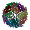
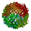
| Entry | Database: PDB / ID: 1ies | ||||||
|---|---|---|---|---|---|---|---|
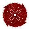
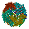
| Title | TETRAGONAL CRYSTAL STRUCTURE OF NATIVE HORSE SPLEEN FERRITIN | ||||||
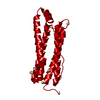
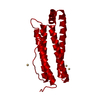
 Components Components | FERRITIN | ||||||
 Keywords Keywords | IRON STORAGE / APOFERRITIN | ||||||
| Function / homology |  Function and homology information: / intracellular sequestering of iron ion / ferric iron binding / ferrous iron binding / iron ion transport / iron ion binding / cytoplasm Function and homology information: / intracellular sequestering of iron ion / ferric iron binding / ferrous iron binding / iron ion transport / iron ion binding / cytoplasmSimilarity search - Function | ||||||
| Biological species |  Equus caballus (horse) Equus caballus (horse) | ||||||
| Method | X-RAY DIFFRACTION / SYNCHROTRON / MOLECULAR REPLACEMENT / Resolution: 2.6 Å | ||||||
 Authors Authors | Granier, T. / Gallois, B. / Dautant, A. / Langlois D'Estaintot, B. / Precigoux, G. | ||||||
 Citation Citation | Journal: Acta Crystallogr.,Sect.D / Year: 1997 Title: Comparison of the structures of the cubic and tetragonal forms of horse-spleen apoferritin. Authors: Granier, T. / Gallois, B. / Dautant, A. / Langlois d'Estaintot, B. / Precigoux, G. #1: Journal: Acta Crystallogr.,Sect.D / Year: 1996Title: Preliminary Results on X-Ray Diffraction Study of the Tetragonal Form of Native Horse Spleen Ferritin Authors: Granier, T. / Gallois, B. / Dautant, A. / Langlois D'Estaintot, B. / Precigoux, G. #2: Journal: Acta Crystallogr.,Sect.D / Year: 1994Title: A Crystallographic Study of Haem Binding to Ferritin Authors: Precigoux, G. / Yariv, J. / Gallois, B. / Dautant, A. / Courseille, C. / Langlois D'Estaintot, B. #3: Journal: Nature / Year: 1980Title: Helix Packing and Subunit Conformation in Horse Spleen Apoferritin Authors: Clegg, G.A. / Stansfield, R.F. / Bourne, P.E. / Harrison, P.M. #4: Journal: J.Mol.Biol. / Year: 1974Title: A Tetragonal Crystal Form of Horse Spleen Apoferritin and its Relation to the Cubic Modification Authors: Hoy, T.G. / Harrison, P.M. / Hoare, R.J. | ||||||
| History |
|
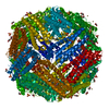
- Structure visualization
| Structure viewer | Molecule: MolmilJmol/JSmol |
|---|
- Downloads & links
-Download
| PDBx/mmCIF format | 1ies.cif.gz | 217.1 KB | Display | PDBx/mmCIF format |
|---|---|---|---|---|
| PDB format | pdb1ies.ent.gz | 177.3 KB | Display | PDB format |
| PDBx/mmJSON format | 1ies.json.gz | Tree view | PDBx/mmJSON format | |
| Others |  Other downloads Other downloads |
-Validation report
| Arichive directory | https://data.pdbj.org/pub/pdb/validation_reports/ie/1iesftp://data.pdbj.org/pub/pdb/validation_reports/ie/1ies | HTTPS FTP |
|---|
-Related structure data
| Related structure data | 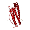 1ierC 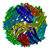 1hrsS S: Starting model for refinement C: citing same article ( |
|---|---|
| Similar structure data |


-Links
 PDBj
PDBj


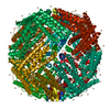
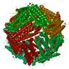
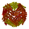
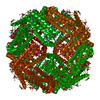
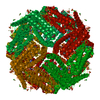
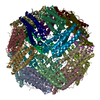
- Assembly
| Deposited unit | 
| |||||||||
|---|---|---|---|---|---|---|---|---|---|---|
| 1 | 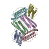
| |||||||||
| Unit cell |
| |||||||||
| Components on special symmetry positions |
|
-Components
| #1: Protein | Mass: 19872.428 Da / Num. of mol.: 6 / Fragment: L-CHAIN / Source method: isolated from a natural source / Source: (natural) Equus caballus (horse) / Organ: SPLEEN / Tissue: SPLEEN / References: UniProt: P02791#2: Chemical | ChemComp-CD /   Mass: 112.411 Da / Num. of mol.: 5 / Source method: obtained synthetically / Formula: Cd Mass: 112.411 Da / Num. of mol.: 5 / Source method: obtained synthetically / Formula: Cd#3: Water | ChemComp-HOH / | Water Mass: 18.015 Da / Num. of mol.: 272 / Source method: isolated from a natural source / Formula: H2O Mass: 18.015 Da / Num. of mol.: 272 / Source method: isolated from a natural source / Formula: H2O |
|---|
-Experimental details
-Experiment
| Experiment | Method: X-RAY DIFFRACTION / Number of used crystals: 2 |
|---|
- Sample preparation
| Crystal | Density Matthews: 3.45 Å3/Da / Density % sol: 64.4 % | ||||||||||||||||||||
|---|---|---|---|---|---|---|---|---|---|---|---|---|---|---|---|---|---|---|---|---|---|
| Crystal | *PLUS | ||||||||||||||||||||
| Crystal grow | *PLUS Method: vapor diffusion, hanging drop | ||||||||||||||||||||
| Components of the solutions | *PLUS
|
-Data collection
| Diffraction | Mean temperature: 293 K |
|---|---|
| Diffraction source | Source: SYNCHROTRON / Site: LURE  / Beamline: D41A / Wavelength: 1.375 / Beamline: D41A / Wavelength: 1.375 |
| Detector | Type: MARRESEARCH / Detector: IMAGE PLATE / Date: Jun 1, 1995 |
| Radiation | Monochromatic (M) / Laue (L): M / Scattering type: x-ray |
| Radiation wavelength | Wavelength: 1.375 Å / Relative weight: 1 |
| Reflection | Resolution: 2.5→15 Å / Num. obs: 51340 / % possible obs: 89.5 % / Observed criterion σ(I): 0 / Redundancy: 6.9 % / Rmerge(I) obs: 0.118 / Net I/σ(I): 19.8 |
| Reflection shell | Resolution: 2.5→2.7 Å / Redundancy: 2.4 % / Rmerge(I) obs: 0.365 / Mean I/σ(I) obs: 1.8 / % possible all: 60.8 |
| Reflection | *PLUS Highest resolution: 2.5 Å / Observed criterion σ(I): 2 / Num. measured all: 354156 |
- Processing
| Software |
| ||||||||||||||||||||||||||||||||||||||||||||||||||||||||||||
|---|---|---|---|---|---|---|---|---|---|---|---|---|---|---|---|---|---|---|---|---|---|---|---|---|---|---|---|---|---|---|---|---|---|---|---|---|---|---|---|---|---|---|---|---|---|---|---|---|---|---|---|---|---|---|---|---|---|---|---|---|---|
| Refinement | Method to determine structure: MOLECULAR REPLACEMENT Starting model: PDB ENTRY 1HRS Resolution: 2.6→8 Å / Data cutoff high absF: 10000000 / Data cutoff low absF: 0 / σ(F): 1 Details: WARNING THE THREE C-TERMINAL RESIDUES 172 - 174, SIDE CHAIN ATOMS FOR RESIDUES SER 1, GLN 3, GLU 45, HIS 49, GLU 53, GLU 56, GLU 57, LYS 67, GLN 82, GLU 136, SER 157 AND GLN 158 ARE POORLY ...Details: WARNING THE THREE C-TERMINAL RESIDUES 172 - 174, SIDE CHAIN ATOMS FOR RESIDUES SER 1, GLN 3, GLU 45, HIS 49, GLU 53, GLU 56, GLU 57, LYS 67, GLN 82, GLU 136, SER 157 AND GLN 158 ARE POORLY DEFINED IN THE ELECTRON DENSITY MAP, PROBABLY BECAUSE OF DISORDER. ALL THE REMAINING RESIDUES HAVE WELL-DEFINED DENSITY.
| ||||||||||||||||||||||||||||||||||||||||||||||||||||||||||||
| Displacement parameters | Biso mean: 25.7 Å2 | ||||||||||||||||||||||||||||||||||||||||||||||||||||||||||||
| Refine analyze | Luzzati coordinate error obs: 0.26 Å | ||||||||||||||||||||||||||||||||||||||||||||||||||||||||||||
| Refinement step | Cycle: LAST / Resolution: 2.6→8 Å
| ||||||||||||||||||||||||||||||||||||||||||||||||||||||||||||
| Refine LS restraints |
| ||||||||||||||||||||||||||||||||||||||||||||||||||||||||||||
| LS refinement shell | Resolution: 2.6→2.66 Å
| ||||||||||||||||||||||||||||||||||||||||||||||||||||||||||||
| Software | *PLUS Name: X-PLOR / Version: 2.1 / Classification: refinement | ||||||||||||||||||||||||||||||||||||||||||||||||||||||||||||
| Refinement | *PLUS Highest resolution: 2.6 Å / Lowest resolution: 8 Å | ||||||||||||||||||||||||||||||||||||||||||||||||||||||||||||
| Solvent computation | *PLUS | ||||||||||||||||||||||||||||||||||||||||||||||||||||||||||||
| Displacement parameters | *PLUS | ||||||||||||||||||||||||||||||||||||||||||||||||||||||||||||
| Refine LS restraints | *PLUS
| ||||||||||||||||||||||||||||||||||||||||||||||||||||||||||||
| LS refinement shell | *PLUS Lowest resolution: 2.7 Å / Rfactor Rwork: 0.27 |
