 Movie
Movie Controller


+ Open data

- Basic information
| Entry | Database: PDB / ID: 1ie3 | |||||||||
|---|---|---|---|---|---|---|---|---|---|---|
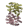
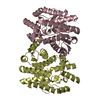
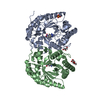
| Title | CRYSTAL STRUCTURE OF R153C E. COLI MALATE DEHYDROGENASE | |||||||||
 Components Components | MALATE DEHYDROGENASE | |||||||||
 Keywords Keywords | OXIDOREDUCTASE / dehydrogenase / malate dehydrogenase / substrate specificity | |||||||||
| Function / homology |  Function and homology informationmalate dehydrogenase activity / fermentation / malate dehydrogenase / L-malate dehydrogenase activity / malate metabolic process / anaerobic respiration / extrinsic component of membrane / tricarboxylic acid cycle / glycolytic process / oxidoreductase activity ...malate dehydrogenase activity / fermentation / malate dehydrogenase / L-malate dehydrogenase activity / malate metabolic process / anaerobic respiration / extrinsic component of membrane / tricarboxylic acid cycle / glycolytic process / oxidoreductase activity / protein homodimerization activity / membrane / cytosol / cytoplasm Function and homology informationmalate dehydrogenase activity / fermentation / malate dehydrogenase / L-malate dehydrogenase activity / malate metabolic process / anaerobic respiration / extrinsic component of membrane / tricarboxylic acid cycle / glycolytic process / oxidoreductase activity ...malate dehydrogenase activity / fermentation / malate dehydrogenase / L-malate dehydrogenase activity / malate metabolic process / anaerobic respiration / extrinsic component of membrane / tricarboxylic acid cycle / glycolytic process / oxidoreductase activity / protein homodimerization activity / membrane / cytosol / cytoplasmSimilarity search - Function | |||||||||
| Biological species |  Escherichia coli (E. coli) Escherichia coli (E. coli) | |||||||||
| Method | X-RAY DIFFRACTION / MOLECULAR REPLACEMENT / Resolution: 2.5 Å | |||||||||
 Authors Authors | Bell, J.K. / Yennawar, H.P. / Wright, S.K. / Thompson, J.R. / Viola, R.E. / Banaszak, L.J. | |||||||||
 Citation Citation | Journal: J.Biol.Chem. / Year: 2001 Title: Structural Analyses of a Malate Dehydrogenase with a Variable Active Site Authors: Bell, J.K. / Yennawar, H.P. / Wright, S.K. / Thompson, J.R. / Viola, R.E. / Banaszak, L.J. #1: Journal: To be PublishedTitle: Alteration of the Specificity of Malate Dehydrogenase by Chemical Modification of an Acitve Site Arginine Authors: Kirk, S.K. / Viola, R.E. | |||||||||
| History |
|
- Structure visualization
| Structure viewer | Molecule: MolmilJmol/JSmol |
|---|
- Downloads & links
-Download
| PDBx/mmCIF format | 1ie3.cif.gz | 238.3 KB | Display | PDBx/mmCIF format |
|---|---|---|---|---|
| PDB format | pdb1ie3.ent.gz | 194.7 KB | Display | PDB format |
| PDBx/mmJSON format | 1ie3.json.gz | Tree view | PDBx/mmJSON format | |
| Others |  Other downloads Other downloads |
-Validation report
| Arichive directory | https://data.pdbj.org/pub/pdb/validation_reports/ie/1ie3ftp://data.pdbj.org/pub/pdb/validation_reports/ie/1ie3 | HTTPS FTP |
|---|
-Related structure data


-Links
 PDBj
PDBj


- Assembly
| Deposited unit | 
| ||||||||
|---|---|---|---|---|---|---|---|---|---|
| 1 | 
| ||||||||
| 2 | 
| ||||||||
| Unit cell |
|
-Components
| #1: Protein | Mass: 32313.141 Da / Num. of mol.: 4 / Mutation: R153C Source method: isolated from a genetically manipulated source Source: (gene. exp.) Escherichia coli (E. coli) / Gene: MDH / Plasmid: pEM6 / Production host: Escherichia coli (E. coli) / References: UniProt: P61889, malate dehydrogenase#2: Chemical | ChemComp-NAD / Nicotinamide adenine dinucleotide  Mass: 663.425 Da / Num. of mol.: 4 / Source method: obtained synthetically / Formula: C21H27N7O14P2 / Comment: NAD*YM Mass: 663.425 Da / Num. of mol.: 4 / Source method: obtained synthetically / Formula: C21H27N7O14P2 / Comment: NAD*YM#3: Chemical | ChemComp-PYR / | Pyruvic acid  Mass: 88.062 Da / Num. of mol.: 1 / Source method: obtained synthetically / Formula: C3H4O3 Mass: 88.062 Da / Num. of mol.: 1 / Source method: obtained synthetically / Formula: C3H4O3#4: Water | ChemComp-HOH / | Water Mass: 18.015 Da / Num. of mol.: 166 / Source method: isolated from a natural source / Formula: H2O Mass: 18.015 Da / Num. of mol.: 166 / Source method: isolated from a natural source / Formula: H2O |
|---|
-Experimental details
-Experiment
| Experiment | Method: X-RAY DIFFRACTION / Number of used crystals: 1 |
|---|
- Sample preparation
| Crystal | Density Matthews: 2.51 Å3/Da / Density % sol: 50.91 % | ||||||||||||||||||||
|---|---|---|---|---|---|---|---|---|---|---|---|---|---|---|---|---|---|---|---|---|---|
| Crystal grow | Temperature: 297 K / Method: vapor diffusion, hanging drop / pH: 6.5 Details: NaCacodylate, PEG 4000, Ammonium Acetate, pH 6.5, VAPOR DIFFUSION, HANGING DROP, temperature 297K | ||||||||||||||||||||
| Crystal grow | *PLUS Temperature: 24 K / Details: used microseeding | ||||||||||||||||||||
| Components of the solutions | *PLUS
|
-Data collection
| Diffraction | Mean temperature: 297 K |
|---|---|
| Diffraction source | Source: ROTATING ANODE / Type: RIGAKU RU200 / Wavelength: 1.5418 Å |
| Detector | Type: RIGAKU RAXIS IV / Detector: IMAGE PLATE / Date: Sep 15, 1999 |
| Radiation | Monochromator: graphite / Protocol: SINGLE WAVELENGTH / Monochromatic (M) / Laue (L): M / Scattering type: x-ray |
| Radiation wavelength | Wavelength: 1.5418 Å / Relative weight: 1 |
| Reflection | Resolution: 2.5→40 Å / Num. all: 95846 / Num. obs: 66535 / % possible obs: 96.4 % / Observed criterion σ(F): 0 / Observed criterion σ(I): 0 / Redundancy: 1.4 % / Biso Wilson estimate: 44.9 Å2 / Rmerge(I) obs: 0.115 / Net I/σ(I): 8.5 |
| Reflection shell | Resolution: 2.5→2.59 Å / Redundancy: 1.4 % / Rmerge(I) obs: 0.361 / % possible all: 91.7 |
| Reflection | *PLUS Lowest resolution: 40 Å / % possible obs: 87.5 % / Num. measured all: 95846 |
| Reflection shell | *PLUS % possible obs: 90 % |
- Processing
| Software |
| |||||||||||||||||||||||||
|---|---|---|---|---|---|---|---|---|---|---|---|---|---|---|---|---|---|---|---|---|---|---|---|---|---|---|
| Refinement | Method to determine structure: MOLECULAR REPLACEMENT / Resolution: 2.5→39.7 Å / Rfactor Rfree error: 0.004 / Data cutoff high absF: 2483345.77 / Data cutoff low absF: 0 / Isotropic thermal model: RESTRAINED / Cross valid method: THROUGHOUT / σ(F): 0 / σ(I): 0 / Stereochemistry target values: Engh and Huber
| |||||||||||||||||||||||||
| Solvent computation | Solvent model: FLAT MODEL / Bsol: 49 Å2 / ksol: 0.302 e/Å3 | |||||||||||||||||||||||||
| Displacement parameters | Biso mean: 44.9 Å2
| |||||||||||||||||||||||||
| Refine analyze |
| |||||||||||||||||||||||||
| Refinement step | Cycle: LAST / Resolution: 2.5→39.7 Å
| |||||||||||||||||||||||||
| Refine LS restraints |
| |||||||||||||||||||||||||
| LS refinement shell | Resolution: 2.5→2.66 Å / Rfactor Rfree error: 0.014 / Total num. of bins used: 6
| |||||||||||||||||||||||||
| Xplor file |
| |||||||||||||||||||||||||
| Software | *PLUS Name: CNS / Version: 0.9 / Classification: refinement | |||||||||||||||||||||||||
| Refinement | *PLUS σ(F): 0 / % reflection Rfree: 10.1 % | |||||||||||||||||||||||||
| Solvent computation | *PLUS | |||||||||||||||||||||||||
| Displacement parameters | *PLUS Biso mean: 44.9 Å2 | |||||||||||||||||||||||||
| Refine LS restraints | *PLUS
| |||||||||||||||||||||||||
| LS refinement shell | *PLUS Rfactor Rfree: 0.252 / % reflection Rfree: 9.7 % / Rfactor Rwork: 0.186 |
