 Movie
Movie Controller

[English] 日本語
 Yorodumi
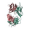
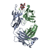
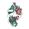
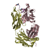
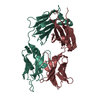
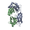
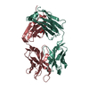
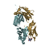
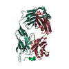
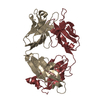
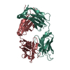
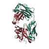
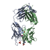
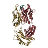
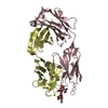
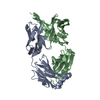
- PDB-1i9i: NATIVE CRYSTAL STRUCTURE OF THE RECOMBINANT MONOCLONAL WILD TYPE ... -
+ Open data

- Basic information
| Entry | Database: PDB / ID: 1i9i | ||||||
|---|---|---|---|---|---|---|---|
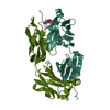
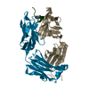
| Title | NATIVE CRYSTAL STRUCTURE OF THE RECOMBINANT MONOCLONAL WILD TYPE ANTI-TESTOSTERONE FAB FRAGMENT | ||||||
 Components Components |
| ||||||
 Keywords Keywords |  IMMUNE SYSTEM / fab fragment / anti-testosterone / recombinant / monoclonal IMMUNE SYSTEM / fab fragment / anti-testosterone / recombinant / monoclonal | ||||||
| Function / homology |  Function and homology information Function and homology informationimmunoglobulin complex, circulating / immunoglobulin receptor binding / complement activation, classical pathway / antigen binding / antibacterial humoral response / blood microparticle / immune response / extracellular space / extracellular exosome / metal ion bindingSimilarity search - Function | ||||||
| Biological species |  Mus musculus (house mouse) Mus musculus (house mouse) | ||||||
| Method | X-RAY DIFFRACTION / MOLECULAR REPLACEMENT / Resolution: 2.72 Å | ||||||
 Authors Authors | Valjakka, J. / Takkinenz, K. / Teerinen, T. / Soderlund, H. / Rouvinen, J. | ||||||
 Citation Citation | Journal: J.Biol.Chem. / Year: 2002 Title: Structural insights into steroid hormone binding: the crystal structure of a recombinant anti-testosterone Fab fragment in free and testosterone-bound forms. Authors: Valjakka, J. / Takkinenz, K. / Teerinen, T. / Soderlund, H. / Rouvinen, J. #1: Journal: Acta Crystallogr.,Sect.D / Year: 2000Title: X-ray studies of recombinant anti-testosterone fab fragments: the use of peg 3350 in crystallization Authors: Valjakka, J. / Hemminki, A. / Teerinen, T. / Takkinen, K. / Rouvinen, J. | ||||||
| History |
|
- Structure visualization
| Structure viewer | Molecule: MolmilJmol/JSmol |
|---|
- Downloads & links
-Download
| PDBx/mmCIF format | 1i9i.cif.gz | 95.5 KB | Display | PDBx/mmCIF format |
|---|---|---|---|---|
| PDB format | pdb1i9i.ent.gz | 73.8 KB | Display | PDB format |
| PDBx/mmJSON format | 1i9i.json.gz | Tree view | PDBx/mmJSON format | |
| Others |  Other downloads Other downloads |
-Validation report
| Arichive directory | https://data.pdbj.org/pub/pdb/validation_reports/i9/1i9iftp://data.pdbj.org/pub/pdb/validation_reports/i9/1i9i | HTTPS FTP |
|---|
-Related structure data


-Links
 PDBj
PDBj

- Assembly
| Deposited unit | 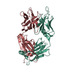
| ||||||||
|---|---|---|---|---|---|---|---|---|---|
| 1 |
| ||||||||
| Unit cell |
|
-Components
| #1: Antibody | Mass: 24056.625 Da / Num. of mol.: 1 Source method: isolated from a genetically manipulated source Source: (gene. exp.) Mus musculus (house mouse) / Plasmid: PKKTAC / Production host:  Escherichia coli (E. coli) / Strain (production host): RV308 / References: UniProt: Q99M37, UniProt: Q65ZC0*PLUS Escherichia coli (E. coli) / Strain (production host): RV308 / References: UniProt: Q99M37, UniProt: Q65ZC0*PLUS |
|---|---|
| #2: Antibody | Mass: 23509.566 Da / Num. of mol.: 1 Source method: isolated from a genetically manipulated source Source: (gene. exp.) Mus musculus (house mouse) / Plasmid: PKKTAC / Production host: Escherichia coli (E. coli) / Strain (production host): RV308 / References: UniProt: Q9R1A4, UniProt: Q91Z05*PLUS |
| #3: Water | ChemComp-HOH / Water Mass: 18.015 Da / Num. of mol.: 88 / Source method: isolated from a natural source / Formula: H2O Mass: 18.015 Da / Num. of mol.: 88 / Source method: isolated from a natural source / Formula: H2O |
-Experimental details
-Experiment
| Experiment | Method: X-RAY DIFFRACTION / Number of used crystals: 1 |
|---|
- Sample preparation
| Crystal | Density Matthews: 2.96 Å3/Da / Density % sol: 58.44 % | ||||||||||||||||||||||||
|---|---|---|---|---|---|---|---|---|---|---|---|---|---|---|---|---|---|---|---|---|---|---|---|---|---|
| Crystal grow | Temperature: 298 K / Method: vapor diffusion, hanging drop / pH: 5.6 Details: peg3350, pH 5.6, VAPOR DIFFUSION, HANGING DROP, temperature 298K | ||||||||||||||||||||||||
| Crystal grow | *PLUS | ||||||||||||||||||||||||
| Components of the solutions | *PLUS
|
-Data collection
| Diffraction | Mean temperature: 120 K |
|---|---|
| Diffraction source | Source: ROTATING ANODE / Type: RIGAKU RU200HB / Wavelength: 1.5418 Å |
| Detector | Type: RIGAKU RAXIS IIC / Detector: IMAGE PLATE / Details: MSC Confocal Blue Optics |
| Radiation | Monochromator: MSC Confocal Blue Optics / Protocol: SINGLE WAVELENGTH / Monochromatic (M) / Laue (L): M / Scattering type: x-ray |
| Radiation wavelength | Wavelength: 1.5418 Å / Relative weight: 1 |
| Reflection | Resolution: 2.72→99 Å / Num. all: 103739 / Num. obs: 15557 / % possible obs: 99.9 % / Observed criterion σ(I): 1 / Redundancy: 6.7 % / Rmerge(I) obs: 0.069 |
| Reflection shell | Resolution: 2.72→2.82 Å / Rmerge(I) obs: 0.304 / % possible all: 99.9 |
| Reflection | *PLUS Lowest resolution: 99 Å / Num. measured all: 103739 |
| Reflection shell | *PLUS % possible obs: 99.9 % / Mean I/σ(I) obs: 4.4 |
- Processing
| Software |
| ||||||||||||||||||||
|---|---|---|---|---|---|---|---|---|---|---|---|---|---|---|---|---|---|---|---|---|---|
| Refinement | Method to determine structure: MOLECULAR REPLACEMENT Starting model: Fab fragment 4-4-20 Resolution: 2.72→99 Å / σ(F): 1
| ||||||||||||||||||||
| Refinement step | Cycle: LAST / Resolution: 2.72→99 Å
| ||||||||||||||||||||
| Refine LS restraints |
| ||||||||||||||||||||
| Xplor file |
| ||||||||||||||||||||
| Refinement | *PLUS Lowest resolution: 500 Å / σ(F): 1 / Rfactor obs: 0.195 | ||||||||||||||||||||
| Solvent computation | *PLUS | ||||||||||||||||||||
| Displacement parameters | *PLUS | ||||||||||||||||||||
| Refine LS restraints | *PLUS
|
