 Movie
Movie Controller

[English] 日本語
 Yorodumi
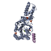
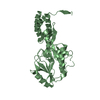
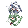
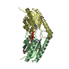
- PDB-1i4w: THE CRYSTAL STRUCTURE OF THE TRANSCRIPTION FACTOR SC-MTTFB OFFERS... -
+ Open data

- Basic information
| Entry | Database: PDB / ID: 1i4w | ||||||
|---|---|---|---|---|---|---|---|
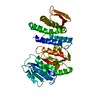
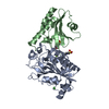
| Title | THE CRYSTAL STRUCTURE OF THE TRANSCRIPTION FACTOR SC-MTTFB OFFERS INTRIGUING INSIGHTS INTO MITOCHONDRIAL TRANSCRIPTION | ||||||
 Components Components | MITOCHONDRIAL REPLICATION PROTEIN MTF1 | ||||||
 Keywords Keywords |  TRANSCRIPTION / MITOCHONDRIAL TRANSCRIPTION FACTOR / TRANSCRIPTION INITIATION TRANSCRIPTION / MITOCHONDRIAL TRANSCRIPTION FACTOR / TRANSCRIPTION INITIATION | ||||||
| Function / homology |  Function and homology information Function and homology informationMitochondrial transcription initiation / mitochondrial DNA-directed RNA polymerase complex / mitochondrial transcription factor activity / transcription initiation at mitochondrial promoter / mitochondrial transcription / positive regulation of DNA-templated transcription, elongation / Transferases; Transferring one-carbon groups; Methyltransferases / methyltransferase activity / mitochondrial intermembrane space / methylation ...Mitochondrial transcription initiation / mitochondrial DNA-directed RNA polymerase complex / mitochondrial transcription factor activity / transcription initiation at mitochondrial promoter / mitochondrial transcription / positive regulation of DNA-templated transcription, elongation / Transferases; Transferring one-carbon groups; Methyltransferases / methyltransferase activity / mitochondrial intermembrane space / methylation / mitochondrial matrix / mitochondrion / DNA binding / RNA bindingSimilarity search - Function | ||||||
| Biological species |  Saccharomyces cerevisiae (brewer's yeast) Saccharomyces cerevisiae (brewer's yeast) | ||||||
| Method | X-RAY DIFFRACTION / SYNCHROTRON / SIRAS / Resolution: 2.6 Å | ||||||
 Authors Authors | Schubot, F.D. / Chen, C.-J. / Rose, J.P. / Dailey, T.A. / Dailey, H.A. / Wang, B.-C. | ||||||
 Citation Citation | Journal: Protein Sci. / Year: 2001 Title: Crystal structure of the transcription factor sc-mtTFB offers insights into mitochondrial transcription. Authors: Schubot, F.D. / Chen, C.J. / Rose, J.P. / Dailey, T.A. / Dailey, H.A. / Wang, B.C. | ||||||
| History |
|
- Structure visualization
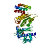
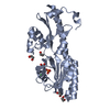
| Structure viewer | Molecule: MolmilJmol/JSmol |
|---|
- Downloads & links
-Download
| PDBx/mmCIF format | 1i4w.cif.gz | 78.8 KB | Display | PDBx/mmCIF format |
|---|---|---|---|---|
| PDB format | pdb1i4w.ent.gz | 58.7 KB | Display | PDB format |
| PDBx/mmJSON format | 1i4w.json.gz | Tree view | PDBx/mmJSON format | |
| Others |  Other downloads Other downloads |
-Validation report
| Arichive directory | https://data.pdbj.org/pub/pdb/validation_reports/i4/1i4wftp://data.pdbj.org/pub/pdb/validation_reports/i4/1i4w | HTTPS FTP |
|---|
-Related structure data
| Similar structure data |
|---|
-Links
 PDBj
PDBj
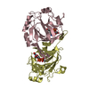
- Assembly
| Deposited unit | 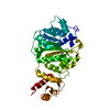
| ||||||||
|---|---|---|---|---|---|---|---|---|---|
| 1 |
| ||||||||
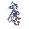
| Unit cell |
|
-Components
| #1: Protein | Mass: 41094.152 Da / Num. of mol.: 1 Source method: isolated from a genetically manipulated source Source: (gene. exp.) Saccharomyces cerevisiae (brewer's yeast)Gene: MTF1 / Plasmid: PTRCHISC / Production host:  Escherichia coli (E. coli) / Strain (production host): JM109 / References: UniProt: P14908 Escherichia coli (E. coli) / Strain (production host): JM109 / References: UniProt: P14908 | ||
|---|---|---|---|
| #2: Chemical | Xenon  Mass: 131.293 Da / Num. of mol.: 3 / Source method: obtained synthetically / Formula: Xe Mass: 131.293 Da / Num. of mol.: 3 / Source method: obtained synthetically / Formula: Xe#3: Water | ChemComp-HOH / | Water Mass: 18.015 Da / Num. of mol.: 21 / Source method: isolated from a natural source / Formula: H2O Mass: 18.015 Da / Num. of mol.: 21 / Source method: isolated from a natural source / Formula: H2O |
-Experimental details
-Experiment
| Experiment | Method: X-RAY DIFFRACTION / Number of used crystals: 1 |
|---|
- Sample preparation
| Crystal | Density Matthews: 2.4 Å3/Da / Density % sol: 48 % | ||||||||||||||||||||||||||||||
|---|---|---|---|---|---|---|---|---|---|---|---|---|---|---|---|---|---|---|---|---|---|---|---|---|---|---|---|---|---|---|---|
| Crystal grow | Temperature: 291 K / Method: vapor diffusion, hanging drop / pH: 6.5 Details: PEG8000, calcium acetate, cacodylate, xylitol, pH 6.5, VAPOR DIFFUSION, HANGING DROP, temperature 291K | ||||||||||||||||||||||||||||||
| Crystal grow | *PLUS pH: 8 / Details: Schubot, G.S., (2000) Acta Crystallogr., D56, 902. | ||||||||||||||||||||||||||||||
| Components of the solutions | *PLUS
|
-Data collection
| Diffraction | Mean temperature: 100 K |
|---|---|
| Diffraction source | Source: SYNCHROTRON / Site: APS  / Beamline: 19-ID / Wavelength: 1 / Wavelength: 1 Å / Beamline: 19-ID / Wavelength: 1 / Wavelength: 1 Å |
| Detector | Type: MARRESEARCH / Detector: CCD / Date: Nov 21, 1999 |
| Radiation | Monochromator: Si 111 CHANNEL / Protocol: SINGLE WAVELENGTH / Monochromatic (M) / Laue (L): M / Scattering type: x-ray |
| Radiation wavelength | Wavelength: 1 Å / Relative weight: 1 |
| Reflection | Resolution: 2.6→20 Å / Num. all: 12000 / Num. obs: 10896 / % possible obs: 90.8 % / Observed criterion σ(F): 0 / Observed criterion σ(I): 0 / Redundancy: 5 % / Biso Wilson estimate: 7.7 Å2 / Rmerge(I) obs: 0.058 / Rsym value: 0.055 / Net I/σ(I): 19.5 |
| Reflection shell | Resolution: 2.6→2.7 Å / Redundancy: 3.2 % / Rmerge(I) obs: 0.17 / Mean I/σ(I) obs: 4.6 / Num. unique all: 852 / Rsym value: 0.2 / % possible all: 63.4 |
| Reflection | *PLUS Num. measured all: 202625 / Rmerge(I) obs: 0.06 |
| Reflection shell | *PLUS % possible obs: 63.4 % |
- Processing
| Software |
| ||||||||||||||||||||||||||||||||||||||||||||||||||||||||||||||||||||||||||||||||
|---|---|---|---|---|---|---|---|---|---|---|---|---|---|---|---|---|---|---|---|---|---|---|---|---|---|---|---|---|---|---|---|---|---|---|---|---|---|---|---|---|---|---|---|---|---|---|---|---|---|---|---|---|---|---|---|---|---|---|---|---|---|---|---|---|---|---|---|---|---|---|---|---|---|---|---|---|---|---|---|---|---|
| Refinement | Method to determine structure: SIRAS / Resolution: 2.6→19.88 Å / Rfactor Rfree error: 0.012 / Data cutoff high absF: 382174.1 / Data cutoff low absF: 0 / Isotropic thermal model: RESTRAINED / Cross valid method: THROUGHOUT / σ(F): 0 / σ(I): 0 / Stereochemistry target values: Engh & Huber
| ||||||||||||||||||||||||||||||||||||||||||||||||||||||||||||||||||||||||||||||||
| Solvent computation | Solvent model: FLAT MODEL / Bsol: 48.07 Å2 / ksol: 0.3669 e/Å3 | ||||||||||||||||||||||||||||||||||||||||||||||||||||||||||||||||||||||||||||||||
| Displacement parameters | Biso mean: 43.3 Å2
| ||||||||||||||||||||||||||||||||||||||||||||||||||||||||||||||||||||||||||||||||
| Refine analyze |
| ||||||||||||||||||||||||||||||||||||||||||||||||||||||||||||||||||||||||||||||||
| Refinement step | Cycle: LAST / Resolution: 2.6→19.88 Å
| ||||||||||||||||||||||||||||||||||||||||||||||||||||||||||||||||||||||||||||||||
| Refine LS restraints |
| ||||||||||||||||||||||||||||||||||||||||||||||||||||||||||||||||||||||||||||||||
| LS refinement shell | Resolution: 2.6→2.76 Å / Rfactor Rfree error: 0.039 / Total num. of bins used: 6
| ||||||||||||||||||||||||||||||||||||||||||||||||||||||||||||||||||||||||||||||||
| Xplor file |
| ||||||||||||||||||||||||||||||||||||||||||||||||||||||||||||||||||||||||||||||||
| Software | *PLUS Name: CNS / Version: 1 / Classification: refinement | ||||||||||||||||||||||||||||||||||||||||||||||||||||||||||||||||||||||||||||||||
| Refinement | *PLUS σ(F): 0 / % reflection Rfree: 5.2 % | ||||||||||||||||||||||||||||||||||||||||||||||||||||||||||||||||||||||||||||||||
| Solvent computation | *PLUS | ||||||||||||||||||||||||||||||||||||||||||||||||||||||||||||||||||||||||||||||||
| Displacement parameters | *PLUS Biso mean: 43.3 Å2 | ||||||||||||||||||||||||||||||||||||||||||||||||||||||||||||||||||||||||||||||||
| Refine LS restraints | *PLUS
| ||||||||||||||||||||||||||||||||||||||||||||||||||||||||||||||||||||||||||||||||
| LS refinement shell | *PLUS Rfactor Rfree: 0.325 / % reflection Rfree: 5.3 % / Rfactor Rwork: 0.255 |
