 Movie
Movie Controller

[English] 日本語
 Yorodumi
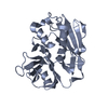
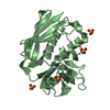
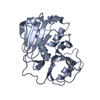
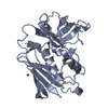
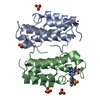
- PDB-1i4g: Crystal structure of Staphylococcal enterotoxin A mutant H187A wi... -
+ Open data

- Basic information
| Entry | Database: PDB / ID: 1i4g | ||||||
|---|---|---|---|---|---|---|---|
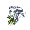
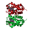
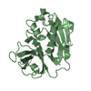
| Title | Crystal structure of Staphylococcal enterotoxin A mutant H187A with reduced Zn2+ affinity | ||||||
 Components Components | ENTEROTOXIN TYPE A | ||||||
 Keywords Keywords |  TOXIN / beta-grasp / beta-barrel TOXIN / beta-grasp / beta-barrel | ||||||
| Function / homology |  Function and homology information Function and homology informationMHC class II protein binding / toxin activity / extracellular region / metal ion bindingSimilarity search - Function | ||||||
| Biological species |   Staphylococcus aureus (bacteria) Staphylococcus aureus (bacteria) | ||||||
| Method | X-RAY DIFFRACTION / MOLECULAR REPLACEMENT / Resolution: 2.1 Å | ||||||
 Authors Authors | Hakansson, M. / Antonsson, P. / Bjork, P. / Svensson, L.A. | ||||||
 Citation Citation | Journal: J.Biol.Inorg.Chem. / Year: 2001 Title: Cooperative zinc binding in a staphylococcal enterotoxin A mutant mimics the SEA-MHC class II interaction Authors: Hakansson, M. / Antonsson, P. / Bjork, P. / Svensson, L.A. | ||||||
| History |
|
- Structure visualization
| Structure viewer | Molecule: MolmilJmol/JSmol |
|---|
- Downloads & links
-Download
| PDBx/mmCIF format | 1i4g.cif.gz | 109.3 KB | Display | PDBx/mmCIF format |
|---|---|---|---|---|
| PDB format | pdb1i4g.ent.gz | 83.4 KB | Display | PDB format |
| PDBx/mmJSON format | 1i4g.json.gz | Tree view | PDBx/mmJSON format | |
| Others |  Other downloads Other downloads |
-Validation report
| Arichive directory | https://data.pdbj.org/pub/pdb/validation_reports/i4/1i4gftp://data.pdbj.org/pub/pdb/validation_reports/i4/1i4g | HTTPS FTP |
|---|
-Related structure data
| Related structure data |  1i4hC  1esfS C: citing same article ( S: Starting model for refinement |
|---|---|
| Similar structure data |

-Links
 PDBj
PDBj
- Assembly
| Deposited unit | 
| ||||||||
|---|---|---|---|---|---|---|---|---|---|
| 1 | 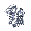
| ||||||||
| 2 | 
| ||||||||
| Unit cell |
|
-Components
| #1: Protein | Mass: 27063.236 Da / Num. of mol.: 2 / Mutation: H187A Source method: isolated from a genetically manipulated source Source: (gene. exp.) Staphylococcus aureus (bacteria) / Plasmid: PKP889 / Production host: Escherichia coli (E. coli) / Strain (production host): UL635 / References: UniProt: P0A0L2#2: Chemical | ChemComp-ZN / |   Mass: 65.409 Da / Num. of mol.: 1 / Source method: obtained synthetically / Formula: Zn Mass: 65.409 Da / Num. of mol.: 1 / Source method: obtained synthetically / Formula: Zn#3: Chemical | ChemComp-SO4 / | Sulfate  Mass: 96.063 Da / Num. of mol.: 1 / Source method: obtained synthetically / Formula: SO4 Mass: 96.063 Da / Num. of mol.: 1 / Source method: obtained synthetically / Formula: SO4#4: Water | ChemComp-HOH / | Water Mass: 18.015 Da / Num. of mol.: 225 / Source method: isolated from a natural source / Formula: H2O Mass: 18.015 Da / Num. of mol.: 225 / Source method: isolated from a natural source / Formula: H2O |
|---|
-Experimental details
-Experiment
| Experiment | Method: X-RAY DIFFRACTION / Number of used crystals: 1 |
|---|
- Sample preparation
| Crystal | Density Matthews: 2.49 Å3/Da / Density % sol: 46 % | ||||||||||||||||||||||||||||||||||||
|---|---|---|---|---|---|---|---|---|---|---|---|---|---|---|---|---|---|---|---|---|---|---|---|---|---|---|---|---|---|---|---|---|---|---|---|---|---|
| Crystal grow | Temperature: 298 K / Method: evaporation / pH: 6 Details: PEG 8000, lithium sulphate, pipes, pH 6.0, EVAPORATION, temperature 298K | ||||||||||||||||||||||||||||||||||||
| Crystal grow | *PLUS pH: 8 / Method: vapor diffusionDetails: drop was mixed with an equal volume of reservoir solution | ||||||||||||||||||||||||||||||||||||
| Components of the solutions | *PLUS
|
-Data collection
| Diffraction | Mean temperature: 298 K |
|---|---|
| Diffraction source | Source: ROTATING ANODE / Type: RIGAKU RU200 / Wavelength: 1.5418 Å |
| Detector | Type: MARRESEARCH / Detector: IMAGE PLATE / Date: Jul 17, 1996 |
| Radiation | Monochromator: graphite / Protocol: SINGLE WAVELENGTH / Monochromatic (M) / Laue (L): M / Scattering type: x-ray |
| Radiation wavelength | Wavelength: 1.5418 Å / Relative weight: 1 |
| Reflection | Resolution: 2.1→20 Å / Num. all: 30860 / Num. obs: 30705 / % possible obs: 95.2 % / Observed criterion σ(F): 0 / Observed criterion σ(I): 0 / Redundancy: 5.1 % / Biso Wilson estimate: 39.6 Å2 / Rmerge(I) obs: 0.07 / Net I/σ(I): 14.2 |
| Reflection shell | Resolution: 2.1→2.3 Å / Redundancy: 3.4 % / Rmerge(I) obs: 0.25 / Mean I/σ(I) obs: 1.6 / Num. unique all: 7022 / % possible all: 92.7 |
| Reflection | *PLUS Num. measured all: 158044 |
| Reflection shell | *PLUS % possible obs: 92.7 % |
- Processing
| Software |
| ||||||||||||||||||||
|---|---|---|---|---|---|---|---|---|---|---|---|---|---|---|---|---|---|---|---|---|---|
| Refinement | Method to determine structure: MOLECULAR REPLACEMENT Starting model: pdb entry 1ESF Resolution: 2.1→20 Å / Isotropic thermal model: Isotropic / Cross valid method: THROUGHOUT / σ(F): 0 / σ(I): 0 / Stereochemistry target values: Engh & Huber
| ||||||||||||||||||||
| Displacement parameters | Biso mean: 39.3 Å2 | ||||||||||||||||||||
| Refine analyze |
| ||||||||||||||||||||
| Refinement step | Cycle: LAST / Resolution: 2.1→20 Å
| ||||||||||||||||||||
| Refine LS restraints |
| ||||||||||||||||||||
| Software | *PLUS Name: X-PLOR / Version: 3.1 / Classification: refinement | ||||||||||||||||||||
| Refine LS restraints | *PLUS
|
