 Movie
Movie Controller


+ Open data

- Basic information
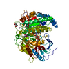
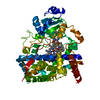
| Entry | Database: PDB / ID: 1i19 | ||||||
|---|---|---|---|---|---|---|---|
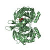
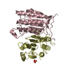
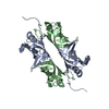
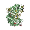
| Title | CRYSTAL STRUCTURE OF CHOLESTEROL OXIDASE FROM B.STEROLICUM | ||||||
 Components Components | CHOLESTEROL OXIDASE | ||||||
 Keywords Keywords | OXIDOREDUCTASE / MIX ALPHA BETA / covalent FAD / Flavoenzyme | ||||||
| Function / homology |  Function and homology information Function and homology informationoxidoreductase activity, acting on the CH-OH group of donors, oxygen as acceptor / FAD binding / metal ion bindingSimilarity search - Function | ||||||
| Biological species |  Brevibacterium sterolicum (bacteria) Brevibacterium sterolicum (bacteria) | ||||||
| Method | X-RAY DIFFRACTION / SYNCHROTRON / MIR / Resolution: 1.7 Å | ||||||
 Authors Authors | Coulombe, R. / Yue, K.Q. / Ghisla, S. / Vrielink, A. | ||||||
 Citation Citation | Journal: J.Biol.Chem. / Year: 2001 Title: Oxygen access to the active site of cholesterol oxidase through a narrow channel is gated by an Arg-Glu pair. Authors: Coulombe, R. / Yue, K.Q. / Ghisla, S. / Vrielink, A. #1: Journal: J.STRUCT.BIOL. / Year: 1996Title: Crystallization and Preliminary X-Ray Analysis of Cholesterol Oxidase from Brevibacterium sterolicum Containing Covalently Bound FAD Authors: Croteau, N. / Vrielink, A. | ||||||
| History |
|
- Structure visualization
| Structure viewer | Molecule: MolmilJmol/JSmol |
|---|
- Downloads & links
-Download
| PDBx/mmCIF format | 1i19.cif.gz | 245.3 KB | Display | PDBx/mmCIF format |
|---|---|---|---|---|
| PDB format | pdb1i19.ent.gz | 202.6 KB | Display | PDB format |
| PDBx/mmJSON format | 1i19.json.gz | Tree view | PDBx/mmJSON format | |
| Others |  Other downloads Other downloads |
-Validation report
| Arichive directory | https://data.pdbj.org/pub/pdb/validation_reports/i1/1i19ftp://data.pdbj.org/pub/pdb/validation_reports/i1/1i19 | HTTPS FTP |
|---|
-Related structure data
| Similar structure data |
|---|
-Links
 PDBj
PDBj


- Assembly
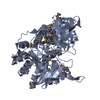
| Deposited unit | 
| ||||||||
|---|---|---|---|---|---|---|---|---|---|
| 1 | 
| ||||||||
| 2 | 
| ||||||||
| Unit cell |
|
-Components
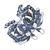
-Protein , 1 types, 2 molecules AB
| #1: Protein | Mass: 61575.082 Da / Num. of mol.: 2 Source method: isolated from a genetically manipulated source Details: COVALENT FAD / Source: (gene. exp.) Brevibacterium sterolicum (bacteria) / Production host: Escherichia coli (E. coli) / References: UniProt: Q7SID9, cholesterol oxidase |
|---|
-Non-polymers , 5 types, 1023 molecules 


| #2: Chemical | ChemComp-EDO / Ethylene glycol Mass: 62.068 Da / Num. of mol.: 12 / Source method: obtained synthetically / Formula: C2H6O2 Mass: 62.068 Da / Num. of mol.: 12 / Source method: obtained synthetically / Formula: C2H6O2#3: Chemical | Flavin adenine dinucleotide Mass: 785.550 Da / Num. of mol.: 2 / Source method: obtained synthetically / Formula: C27H33N9O15P2 / Comment: FAD*YM Mass: 785.550 Da / Num. of mol.: 2 / Source method: obtained synthetically / Formula: C27H33N9O15P2 / Comment: FAD*YM#4: Chemical |  Mass: 54.938 Da / Num. of mol.: 3 / Source method: obtained synthetically / Formula: Mn Mass: 54.938 Da / Num. of mol.: 3 / Source method: obtained synthetically / Formula: Mn#5: Chemical | ChemComp-CAC / | Cacodylic acid Mass: 136.989 Da / Num. of mol.: 1 / Source method: obtained synthetically / Formula: C2H6AsO2 Mass: 136.989 Da / Num. of mol.: 1 / Source method: obtained synthetically / Formula: C2H6AsO2#6: Water | ChemComp-HOH / | WaterMass: 18.015 Da / Num. of mol.: 1005 / Source method: isolated from a natural source / Formula: H2O |
|---|
-Experimental details
-Experiment
| Experiment | Method: X-RAY DIFFRACTION / Number of used crystals: 1 |
|---|
- Sample preparation
| Crystal | Density Matthews: 2.99 Å3/Da / Density % sol: 58.81 % | ||||||||||||||||||||||||||||||||||||
|---|---|---|---|---|---|---|---|---|---|---|---|---|---|---|---|---|---|---|---|---|---|---|---|---|---|---|---|---|---|---|---|---|---|---|---|---|---|
| Crystal grow | Temperature: 290 K / Method: vapor diffusion, hanging drop / pH: 5.2 Details: PEG 8K, MnSO4, NaCacodylate, pH 5.2, VAPOR DIFFUSION, HANGING DROP, temperature 290K | ||||||||||||||||||||||||||||||||||||
| Crystal grow | *PLUS Temperature: 22 ℃ / pH: 7 / Details: Croteau, N., (1996) J.STRUCT.BIOL., 116, 317. | ||||||||||||||||||||||||||||||||||||
| Components of the solutions | *PLUS
|
-Data collection
| Diffraction | Mean temperature: 100 K |
|---|---|
| Diffraction source | Source: SYNCHROTRON / Site: NSLS  / Beamline: X8C / Wavelength: 0.979 Å / Beamline: X8C / Wavelength: 0.979 Å |
| Detector | Type: ADSC QUANTUM 4 / Detector: CCD / Date: Aug 27, 1999 |
| Radiation | Monochromator: crystals Si(111) / Protocol: SINGLE WAVELENGTH / Monochromatic (M) / Laue (L): M / Scattering type: x-ray |
| Radiation wavelength | Wavelength: 0.979 Å / Relative weight: 1 |
| Reflection | Resolution: 1.7→100 Å / Num. all: 554010 / Num. obs: 550182 / % possible obs: 94.8 % / Observed criterion σ(I): 1 / Redundancy: 3.7 % / Biso Wilson estimate: 17.8 Å2 / Rmerge(I) obs: 0.065 / Net I/σ(I): 17.6 |
| Reflection shell | Resolution: 1.7→1.74 Å / Redundancy: 3.5 % / Rmerge(I) obs: 0.377 / Mean I/σ(I) obs: 2.7 / Num. unique all: 7196 / % possible all: 68.4 |
| Reflection | *PLUS Num. obs: 150202 / Num. measured all: 550182 |
| Reflection shell | *PLUS % possible obs: 68.4 % |
- Processing
| Software |
| ||||||||||||||||||||||||||||||||||||||||||||||||||||||||||||||||||||||||||||||||
|---|---|---|---|---|---|---|---|---|---|---|---|---|---|---|---|---|---|---|---|---|---|---|---|---|---|---|---|---|---|---|---|---|---|---|---|---|---|---|---|---|---|---|---|---|---|---|---|---|---|---|---|---|---|---|---|---|---|---|---|---|---|---|---|---|---|---|---|---|---|---|---|---|---|---|---|---|---|---|---|---|---|
| Refinement | Method to determine structure: MIR / Resolution: 1.7→36.91 Å / Rfactor Rfree error: 0.002 / Data cutoff high absF: 1351706.66 / Data cutoff low absF: 0 / Isotropic thermal model: RESTRAINED / Cross valid method: THROUGHOUT / σ(F): 0 / Stereochemistry target values: Engh & Huber Details: The following atoms have occupancies set to 0.00 or 0.50 because of weak electron density: CG1 and CG2 of VAL 349 chains A and B have occupancy set to 0.00; CG, OD1, ND2 of ASN A 65, all ...Details: The following atoms have occupancies set to 0.00 or 0.50 because of weak electron density: CG1 and CG2 of VAL 349 chains A and B have occupancy set to 0.00; CG, OD1, ND2 of ASN A 65, all atoms of PRO A 330, CG, OD1, OD2 of ASP A 544, CG, OD1, ND2 of ASN B 65, and CG, OD1, OD2 of ASP B 179 have occupancy set to 0.50.
| ||||||||||||||||||||||||||||||||||||||||||||||||||||||||||||||||||||||||||||||||
| Solvent computation | Solvent model: FLAT MODEL / Bsol: 45.73 Å2 / ksol: 0.368 e/Å3 | ||||||||||||||||||||||||||||||||||||||||||||||||||||||||||||||||||||||||||||||||
| Displacement parameters | Biso mean: 19.8 Å2
| ||||||||||||||||||||||||||||||||||||||||||||||||||||||||||||||||||||||||||||||||
| Refine analyze |
| ||||||||||||||||||||||||||||||||||||||||||||||||||||||||||||||||||||||||||||||||
| Refinement step | Cycle: LAST / Resolution: 1.7→36.91 Å
| ||||||||||||||||||||||||||||||||||||||||||||||||||||||||||||||||||||||||||||||||
| Refine LS restraints |
| ||||||||||||||||||||||||||||||||||||||||||||||||||||||||||||||||||||||||||||||||
| LS refinement shell | Resolution: 1.7→1.81 Å / Rfactor Rfree error: 0.006 / Total num. of bins used: 6
| ||||||||||||||||||||||||||||||||||||||||||||||||||||||||||||||||||||||||||||||||
| Xplor file |
| ||||||||||||||||||||||||||||||||||||||||||||||||||||||||||||||||||||||||||||||||
| Software | *PLUS Name: CNS / Version: 0.9 / Classification: refinement | ||||||||||||||||||||||||||||||||||||||||||||||||||||||||||||||||||||||||||||||||
| Refinement | *PLUS σ(F): 0 / % reflection Rfree: 10 % / Rfactor obs: 0.182 | ||||||||||||||||||||||||||||||||||||||||||||||||||||||||||||||||||||||||||||||||
| Solvent computation | *PLUS | ||||||||||||||||||||||||||||||||||||||||||||||||||||||||||||||||||||||||||||||||
| Displacement parameters | *PLUS Biso mean: 19.8 Å2 | ||||||||||||||||||||||||||||||||||||||||||||||||||||||||||||||||||||||||||||||||
| Refine LS restraints | *PLUS
| ||||||||||||||||||||||||||||||||||||||||||||||||||||||||||||||||||||||||||||||||
| LS refinement shell | *PLUS Rfactor Rfree: 0.264 / % reflection Rfree: 10 % / Rfactor Rwork: 0.244 |
