 Movie
Movie Controller

[English] 日本語
 Yorodumi
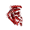
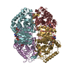
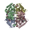
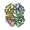
- PDB-1i0z: HUMAN HEART L-LACTATE DEHYDROGENASE H CHAIN, TERNARY COMPLEX WITH... -
+ Open data

- Basic information
| Entry | Database: PDB / ID: 1i0z | ||||||
|---|---|---|---|---|---|---|---|
| Title | HUMAN HEART L-LACTATE DEHYDROGENASE H CHAIN, TERNARY COMPLEX WITH NADH AND OXAMATE | ||||||
 Components Components | L-LACTATE DEHYDROGENASE H CHAIN | ||||||
 Keywords Keywords |  OXIDOREDUCTASE / DEHYDROGENASE / ROSSMANN FOLD OXIDOREDUCTASE / DEHYDROGENASE / ROSSMANN FOLD | ||||||
| Function / homology |  Function and homology informationoxidoreductase complex / lactate metabolic process / Pyruvate metabolism / L-lactate dehydrogenase / L-lactate dehydrogenase activity / NAD metabolic process / pyruvate metabolic process / kinase binding / NAD binding / mitochondrial inner membrane ...oxidoreductase complex / lactate metabolic process / Pyruvate metabolism / L-lactate dehydrogenase / L-lactate dehydrogenase activity / NAD metabolic process / pyruvate metabolic process / kinase binding / NAD binding / mitochondrial inner membrane / membrane raft / mitochondrion / extracellular exosome / membrane / identical protein binding / cytosol / cytoplasm Function and homology informationoxidoreductase complex / lactate metabolic process / Pyruvate metabolism / L-lactate dehydrogenase / L-lactate dehydrogenase activity / NAD metabolic process / pyruvate metabolic process / kinase binding / NAD binding / mitochondrial inner membrane ...oxidoreductase complex / lactate metabolic process / Pyruvate metabolism / L-lactate dehydrogenase / L-lactate dehydrogenase activity / NAD metabolic process / pyruvate metabolic process / kinase binding / NAD binding / mitochondrial inner membrane / membrane raft / mitochondrion / extracellular exosome / membrane / identical protein binding / cytosol / cytoplasmSimilarity search - Function | ||||||
| Biological species |  Homo sapiens (human) Homo sapiens (human) | ||||||
| Method | X-RAY DIFFRACTION / SYNCHROTRON / MOLECULAR REPLACEMENT / Resolution: 2.1 Å | ||||||
 Authors Authors | Read, J.A. / Winter, V.J. / Eszes, C.M. / Sessions, R.B. / Brady, R.L. | ||||||
 Citation Citation | Journal: Proteins / Year: 2001 Title: Structural basis for altered activity of M- and H-isozyme forms of human lactate dehydrogenase. Authors: Read, J.A. / Winter, V.J. / Eszes, C.M. / Sessions, R.B. / Brady, R.L. | ||||||
| History |
|
- Structure visualization
| Structure viewer | Molecule: MolmilJmol/JSmol |
|---|
- Downloads & links
-Download
| PDBx/mmCIF format | 1i0z.cif.gz | 144.7 KB | Display | PDBx/mmCIF format |
|---|---|---|---|---|
| PDB format | pdb1i0z.ent.gz | 114.5 KB | Display | PDB format |
| PDBx/mmJSON format | 1i0z.json.gz | Tree view | PDBx/mmJSON format | |
| Others |  Other downloads Other downloads |
-Validation report
| Arichive directory | https://data.pdbj.org/pub/pdb/validation_reports/i0/1i0zftp://data.pdbj.org/pub/pdb/validation_reports/i0/1i0z | HTTPS FTP |
|---|
-Related structure data
| Related structure data |  1i10C  5ldhS C: citing same article ( S: Starting model for refinement |
|---|---|
| Similar structure data |


-Links
 PDBj
PDBj


- Assembly
| Deposited unit | 
| ||||||||
|---|---|---|---|---|---|---|---|---|---|
| 1 | 
| ||||||||
| Unit cell |
| ||||||||
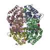
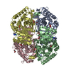
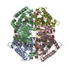
| Details | The biological assembly is a tetramer generated from the dimer in the asymmetric unit |
-Components
| #1: Protein | Mass: 36548.207 Da / Num. of mol.: 2 Source method: isolated from a genetically manipulated source Source: (gene. exp.) Homo sapiens (human) / Gene: LDHB / Organ: HEART / Plasmid: PKK223-3 / Species (production host): Escherichia coli / Production host:  Escherichia coli BL21 (bacteria) / Strain (production host): BL21 / References: UniProt: P07195, L-lactate dehydrogenase Escherichia coli BL21 (bacteria) / Strain (production host): BL21 / References: UniProt: P07195, L-lactate dehydrogenase#2: Chemical | Nicotinamide adenine dinucleotide  Mass: 665.441 Da / Num. of mol.: 2 / Source method: obtained synthetically / Formula: C21H29N7O14P2 Mass: 665.441 Da / Num. of mol.: 2 / Source method: obtained synthetically / Formula: C21H29N7O14P2#3: Chemical | Oxamic acid  Mass: 89.050 Da / Num. of mol.: 2 / Source method: obtained synthetically / Formula: C2H3NO3 Mass: 89.050 Da / Num. of mol.: 2 / Source method: obtained synthetically / Formula: C2H3NO3#4: Water | ChemComp-HOH / | Water Mass: 18.015 Da / Num. of mol.: 318 / Source method: isolated from a natural source / Formula: H2O Mass: 18.015 Da / Num. of mol.: 318 / Source method: isolated from a natural source / Formula: H2O |
|---|
-Experimental details
-Experiment
| Experiment | Method: X-RAY DIFFRACTION / Number of used crystals: 1 |
|---|
- Sample preparation
| Crystal | Density Matthews: 2.27 Å3/Da / Density % sol: 45.74 % | ||||||||||||||||||||||||||||||||||||||||
|---|---|---|---|---|---|---|---|---|---|---|---|---|---|---|---|---|---|---|---|---|---|---|---|---|---|---|---|---|---|---|---|---|---|---|---|---|---|---|---|---|---|
| Crystal grow | Temperature: 291 K / Method: vapor diffusion, hanging drop / pH: 7.5 Details: protein solution: 15 mg/ml H4-ldh, 50 mM Na-Hepes pH 7.5, 2.5 mM NADH, 1 mM oxamate. Well solution: 21% PEG 8k, 100 mM Na-HEPES ph 7.5., VAPOR DIFFUSION, HANGING DROP, temperature 291K | ||||||||||||||||||||||||||||||||||||||||
| Crystal grow | *PLUS Temperature: 18 ℃Details: drop consists of equal amounts of protein and reservoir solutions | ||||||||||||||||||||||||||||||||||||||||
| Components of the solutions | *PLUS
|
-Data collection
| Diffraction | Mean temperature: 100 K |
|---|---|
| Diffraction source | Source: SYNCHROTRON / Site: SRS  / Beamline: PX7.2 / Wavelength: 1.488 Å / Beamline: PX7.2 / Wavelength: 1.488 Å |
| Detector | Type: MARRESEARCH / Detector: IMAGE PLATE / Date: Oct 7, 1999 |
| Radiation | Monochromator: platinum mirrors/Ge 111 monochromator / Protocol: SINGLE WAVELENGTH / Monochromatic (M) / Laue (L): M / Scattering type: x-ray |
| Radiation wavelength | Wavelength: 1.488 Å / Relative weight: 1 |
| Reflection | Resolution: 2.1→30 Å / Num. all: 39237 / Num. obs: 37680 / % possible obs: 96 % / Observed criterion σ(F): 1 / Observed criterion σ(I): 1 / Redundancy: 3.31 % / Rmerge(I) obs: 0.05 / Net I/σ(I): 21.25 |
| Reflection shell | Resolution: 2.1→2.2 Å / Redundancy: 3.13 % / Rmerge(I) obs: 0.114 / Mean I/σ(I) obs: 8.5 / Num. unique all: 4400 / % possible all: 91.1 |
| Reflection shell | *PLUS % possible obs: 91.1 % |
- Processing
| Software |
| ||||||||||||||||||||||||||||
|---|---|---|---|---|---|---|---|---|---|---|---|---|---|---|---|---|---|---|---|---|---|---|---|---|---|---|---|---|---|
| Refinement | Method to determine structure: MOLECULAR REPLACEMENT Starting model: 5ldh Resolution: 2.1→20 Å / SU B: 4.848 / SU ML: 0.133 / Cross valid method: THROUGHOUT / σ(F): 0 / σ(I): 0 / ESU R: 0.247 / ESU R Free: 0.197 / Stereochemistry target values: Engh & Huber Details: METHOD USED FOR BULK SOLVENT MODELLING: BABINET MODEL WITH MASK. PARAMETERS FOR MASK CALCULATION: VDW PROBE RADIUS = 1.40, ION PROBE RADIUS = 0.80, SHRINKAGE RADIUS = 0.80.
| ||||||||||||||||||||||||||||
| Displacement parameters | Biso mean: 19.533 Å2
| ||||||||||||||||||||||||||||
| Refinement step | Cycle: LAST / Resolution: 2.1→20 Å
| ||||||||||||||||||||||||||||
| Refine LS restraints |
| ||||||||||||||||||||||||||||
| LS refinement shell | Resolution: 2.1→2.154 Å / Total num. of bins used: 20 /
| ||||||||||||||||||||||||||||
| Software | *PLUS Name: REFMAC / Classification: refinement | ||||||||||||||||||||||||||||
| Refinement | *PLUS Rfactor obs: 0.179 / Rfactor Rfree: 0.23 / Rfactor Rwork: 0.177 | ||||||||||||||||||||||||||||
| Solvent computation | *PLUS | ||||||||||||||||||||||||||||
| Displacement parameters | *PLUS |
