 Movie
Movie Controller

[English] 日本語
 Yorodumi
- PDB-1hv8: CRYSTAL STRUCTURE OF A DEAD BOX PROTEIN FROM THE HYPERTHERMOPHILE... -
+ Open data

- Basic information
| Entry | Database: PDB / ID: 1hv8 | ||||||
|---|---|---|---|---|---|---|---|
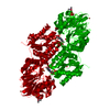
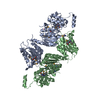
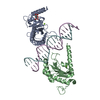
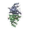
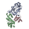
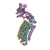
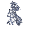
| Title | CRYSTAL STRUCTURE OF A DEAD BOX PROTEIN FROM THE HYPERTHERMOPHILE METHANOCOCCUS JANNASCHII | ||||||
 Components Components | PUTATIVE ATP-DEPENDENT RNA HELICASE MJ0669 | ||||||
 Keywords Keywords |  RNA BINDING PROTEIN / Helicase / RNA-binding Protein / ATPase RNA BINDING PROTEIN / Helicase / RNA-binding Protein / ATPase | ||||||
| Function / homology |  Function and homology information Function and homology information | ||||||
| Biological species |   Methanocaldococcus jannaschii (archaea) Methanocaldococcus jannaschii (archaea) | ||||||
| Method | X-RAY DIFFRACTION / SYNCHROTRON / MAD / Resolution: 3 Å | ||||||
 Authors Authors | Story, R.M. / Li, H. / Abelson, J.N. | ||||||
 Citation Citation | Journal: Proc.Natl.Acad.Sci.USA / Year: 2001 Title: Crystal structure of a DEAD box protein from the hyperthermophile Methanococcus jannaschii. Authors: Story, R.M. / Li, H. / Abelson, J.N. | ||||||
| History |
|
- Structure visualization
| Structure viewer | Molecule: MolmilJmol/JSmol |
|---|
- Downloads & links
-Download
| PDBx/mmCIF format | 1hv8.cif.gz | 136.9 KB | Display | PDBx/mmCIF format |
|---|---|---|---|---|
| PDB format | pdb1hv8.ent.gz | 117.3 KB | Display | PDB format |
| PDBx/mmJSON format | 1hv8.json.gz | Tree view | PDBx/mmJSON format | |
| Others |  Other downloads Other downloads |
-Validation report
| Arichive directory | https://data.pdbj.org/pub/pdb/validation_reports/hv/1hv8ftp://data.pdbj.org/pub/pdb/validation_reports/hv/1hv8 | HTTPS FTP |
|---|
-Related structure data
| Similar structure data |
|---|
-Links
 PDBj
PDBj- Assembly
| Deposited unit | 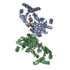
| ||||||||
|---|---|---|---|---|---|---|---|---|---|
| 1 |
| ||||||||
| Unit cell |
|
-Components
| #1: Protein | Mass: 42503.000 Da / Num. of mol.: 2 Source method: isolated from a genetically manipulated source Source: (gene. exp.) Methanocaldococcus jannaschii (archaea)Production host:  Escherichia coli (E. coli) / References: UniProt: Q58083 Escherichia coli (E. coli) / References: UniProt: Q58083#2: Chemical | ChemComp-SO4 / Sulfate  Mass: 96.063 Da / Num. of mol.: 6 / Source method: obtained synthetically / Formula: SO4 Mass: 96.063 Da / Num. of mol.: 6 / Source method: obtained synthetically / Formula: SO4 |
|---|
-Experimental details
-Experiment
| Experiment | Method: X-RAY DIFFRACTION / Number of used crystals: 1 |
|---|
- Sample preparation
| Crystal | Density Matthews: 2.78 Å3/Da / Density % sol: 55.76 % | ||||||||||||||||||||||||||||||||||||||||||||||||||||||||||||
|---|---|---|---|---|---|---|---|---|---|---|---|---|---|---|---|---|---|---|---|---|---|---|---|---|---|---|---|---|---|---|---|---|---|---|---|---|---|---|---|---|---|---|---|---|---|---|---|---|---|---|---|---|---|---|---|---|---|---|---|---|---|
| Crystal grow | Temperature: 303 K / Method: vapor diffusion, sitting drop / pH: 6 Details: ammonium sulfate, acetate,tris, DTT, EDTA, NaN3, pH 6.0, VAPOR DIFFUSION, SITTING DROP, temperature 303K | ||||||||||||||||||||||||||||||||||||||||||||||||||||||||||||
| Crystal grow | *PLUS pH: 7.5 / Method: vapor diffusion | ||||||||||||||||||||||||||||||||||||||||||||||||||||||||||||
| Components of the solutions | *PLUS
|
-Data collection
| Diffraction | Mean temperature: 100 K | ||||||||||||
|---|---|---|---|---|---|---|---|---|---|---|---|---|---|
| Diffraction source | Source: SYNCHROTRON / Site: ALS  / Beamline: 5.0.2 / Wavelength: 0.9795, 0.9793, 0.9611 / Beamline: 5.0.2 / Wavelength: 0.9795, 0.9793, 0.9611 | ||||||||||||
| Detector | Type: ADSC QUANTUM 4 / Detector: CCD / Date: Dec 2, 1999 | ||||||||||||
| Radiation | Monochromator: double crystal / Protocol: MAD / Monochromatic (M) / Laue (L): M / Scattering type: x-ray | ||||||||||||
| Radiation wavelength |
| ||||||||||||
| Reflection | Resolution: 2.95→25 Å / Num. all: 20100 / Num. obs: 20100 / % possible obs: 100 % / Observed criterion σ(I): 3 / Redundancy: 16.1 % / Biso Wilson estimate: 86.2 Å2 / Rmerge(I) obs: 0.097 / Net I/σ(I): 8.9 | ||||||||||||
| Reflection shell | Resolution: 2.95→3 Å / Redundancy: 2 % / Rmerge(I) obs: 0.419 / % possible all: 100 | ||||||||||||
| Reflection | *PLUS % possible obs: 100 % / Num. measured all: 322686 | ||||||||||||
| Reflection shell | *PLUS % possible obs: 100 % |
- Processing
| Software |
| ||||||||||||||||||||
|---|---|---|---|---|---|---|---|---|---|---|---|---|---|---|---|---|---|---|---|---|---|
| Refinement | Method to determine structure: MAD / Resolution: 3→20 Å / σ(F): 0 / Stereochemistry target values: Engh and Huber Details: Residues 299-306 and 330-337 lie in very weak electron density and their structures are tentative
| ||||||||||||||||||||
| Refinement step | Cycle: LAST / Resolution: 3→20 Å
| ||||||||||||||||||||
| Refine LS restraints |
| ||||||||||||||||||||
| Software | *PLUS Name: CNS / Classification: refinement | ||||||||||||||||||||
| Refinement | *PLUS Highest resolution: 3 Å / Lowest resolution: 20 Å / σ(F): 0 / % reflection Rfree: 10 % / Rfactor obs: 0.27 / Rfactor Rwork: 0.27 | ||||||||||||||||||||
| Solvent computation | *PLUS | ||||||||||||||||||||
| Displacement parameters | *PLUS | ||||||||||||||||||||
| Refine LS restraints | *PLUS Type: c_angle_deg / Dev ideal: 1.4 |
