 Movie
Movie Controller

[English] 日本語
 Yorodumi
- PDB-1hux: CRYSTAL STRUCTURE OF THE ACIDAMINOCOCCUS FERMENTANS (R)-2-HYDROXY... -
+ Open data

- Basic information
| Entry | Database: PDB / ID: 1hux | ||||||
|---|---|---|---|---|---|---|---|
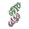
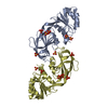
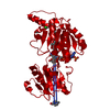
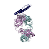
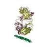
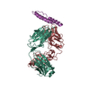
| Title | CRYSTAL STRUCTURE OF THE ACIDAMINOCOCCUS FERMENTANS (R)-2-HYDROXYGLUTARYL-COA DEHYDRATASE COMPONENT A | ||||||
 Components Components | ACTIVATOR OF (R)-2-HYDROXYGLUTARYL-COA DEHYDRATASE | ||||||
 Keywords Keywords | METAL BINDING PROTEIN / actin fold | ||||||
| Function / homology |  Function and homology information Function and homology informationglutamate catabolic process via 2-hydroxyglutarate /  Hydrolases; Acting on acid anhydrides; In phosphorus-containing anhydrides / 4 iron, 4 sulfur cluster binding / hydrolase activity / ATP binding / metal ion binding Hydrolases; Acting on acid anhydrides; In phosphorus-containing anhydrides / 4 iron, 4 sulfur cluster binding / hydrolase activity / ATP binding / metal ion bindingSimilarity search - Function | ||||||
| Biological species |  Acidaminococcus fermentans (bacteria) Acidaminococcus fermentans (bacteria) | ||||||
| Method | X-RAY DIFFRACTION / SYNCHROTRON / MAD / Resolution: 3 Å | ||||||
 Authors Authors | Locher, K.P. / Hans, M. / Yeh, A.P. / Schmid, B. / Buckel, W. / Rees, D.C. | ||||||
 Citation Citation | Journal: J.Mol.Biol. / Year: 2001 Title: Crystal structure of the Acidaminococcus fermentans 2-hydroxyglutaryl-CoA dehydratase component A. Authors: Locher, K.P. / Hans, M. / Yeh, A.P. / Schmid, B. / Buckel, W. / Rees, D.C. | ||||||
| History |
| ||||||
| Remark 999 | SEQUENCE THE TEN ADDITIONAL RESIDUES AT THE C-TERMINUS CORRESPOND TO AN ENGINEERED STREP-II TAG ...SEQUENCE THE TEN ADDITIONAL RESIDUES AT THE C-TERMINUS CORRESPOND TO AN ENGINEERED STREP-II TAG THAT APPEARS DISORDERED IN THE STRUCTURE. |
- Structure visualization
| Structure viewer | Molecule: MolmilJmol/JSmol |
|---|
- Downloads & links
-Download
| PDBx/mmCIF format | 1hux.cif.gz | 97.6 KB | Display | PDBx/mmCIF format |
|---|---|---|---|---|
| PDB format | pdb1hux.ent.gz | 76.8 KB | Display | PDB format |
| PDBx/mmJSON format | 1hux.json.gz | Tree view | PDBx/mmJSON format | |
| Others |  Other downloads Other downloads |
-Validation report
| Arichive directory | https://data.pdbj.org/pub/pdb/validation_reports/hu/1huxftp://data.pdbj.org/pub/pdb/validation_reports/hu/1hux | HTTPS FTP |
|---|
-Related structure data
| Similar structure data |
|---|


-Links
 PDBj
PDBj- Assembly
| Deposited unit | 
| ||||||||
|---|---|---|---|---|---|---|---|---|---|
| 1 |
| ||||||||
| Unit cell |
| ||||||||
| Details | Asymmetric unit contains the functional homodimer with the bridging [4Fe-4S] cluster |
-Components
| #1: Protein | Mass: 28501.736 Da / Num. of mol.: 2 Source method: isolated from a genetically manipulated source Source: (gene. exp.) Acidaminococcus fermentans (bacteria) / Gene: HGDC / Plasmid: PMH6 / Production host: Escherichia coli (E. coli) / Strain (production host): XL-1 BLUE / References: UniProt: P11568#2: Chemical | ChemComp-SF4 / | Iron–sulfur cluster  Mass: 351.640 Da / Num. of mol.: 1 / Source method: obtained synthetically / Formula: Fe4S4 Mass: 351.640 Da / Num. of mol.: 1 / Source method: obtained synthetically / Formula: Fe4S4#3: Chemical | Adenosine diphosphate  Mass: 427.201 Da / Num. of mol.: 2 / Source method: obtained synthetically / Formula: C10H15N5O10P2 / Comment: ADP, energy-carrying molecule*YM Mass: 427.201 Da / Num. of mol.: 2 / Source method: obtained synthetically / Formula: C10H15N5O10P2 / Comment: ADP, energy-carrying molecule*YM |
|---|
-Experimental details
-Experiment
| Experiment | Method: X-RAY DIFFRACTION / Number of used crystals: 1 |
|---|
- Sample preparation
| Crystal | Density Matthews: 2.45 Å3/Da / Density % sol: 49.78 % | ||||||||||||||||||||||||||||||||||||||||||||||||||||||
|---|---|---|---|---|---|---|---|---|---|---|---|---|---|---|---|---|---|---|---|---|---|---|---|---|---|---|---|---|---|---|---|---|---|---|---|---|---|---|---|---|---|---|---|---|---|---|---|---|---|---|---|---|---|---|---|
| Crystal grow | Temperature: 298 K / Method: vapor diffusion, sitting drop / pH: 5.6 Details: 100 mM sodium citrate pH 5.6, 20% PEG 4000, 20% isopropanol, VAPOR DIFFUSION, SITTING DROP, temperature 298.0K | ||||||||||||||||||||||||||||||||||||||||||||||||||||||
| Crystal grow | *PLUS pH: 8 | ||||||||||||||||||||||||||||||||||||||||||||||||||||||
| Components of the solutions | *PLUS
|
-Data collection
| Diffraction | Mean temperature: 130 K | |||||||||||||||
|---|---|---|---|---|---|---|---|---|---|---|---|---|---|---|---|---|
| Diffraction source | Source: SYNCHROTRON / Site: SSRL  / Beamline: BL9-2 / Wavelength: 1.6920, 1.7395, 1.7419, 1.7968 / Beamline: BL9-2 / Wavelength: 1.6920, 1.7395, 1.7419, 1.7968 | |||||||||||||||
| Detector | Type: ADSC QUANTUM 4 / Detector: CCD / Date: Jun 24, 1999 | |||||||||||||||
| Radiation | Monochromator: Double Crystal Si(220) / Protocol: MAD / Monochromatic (M) / Laue (L): M / Scattering type: x-ray | |||||||||||||||
| Radiation wavelength |
| |||||||||||||||
| Reflection | Resolution: 3→43 Å / Num. all: 11525 / Num. obs: 11525 / % possible obs: 99.8 % / Observed criterion σ(I): 3 / Redundancy: 6.92 % / Biso Wilson estimate: 56.32 Å2 / Rmerge(I) obs: 0.084 / Net I/σ(I): 21.7 | |||||||||||||||
| Reflection shell | Resolution: 3→3.05 Å / Redundancy: 6.4 % / Rmerge(I) obs: 0.203 / Mean I/σ(I) obs: 10 / Rsym value: 0.203 / % possible all: 100 | |||||||||||||||
| Reflection | *PLUS Num. measured all: 79725 | |||||||||||||||
| Reflection shell | *PLUS % possible obs: 100 % |
- Processing
| Software |
| ||||||||||||||||||||
|---|---|---|---|---|---|---|---|---|---|---|---|---|---|---|---|---|---|---|---|---|---|
| Refinement | Method to determine structure: MAD / Resolution: 3→50 Å / σ(F): 0 / σ(I): 0 / Stereochemistry target values: Engh & Huber (CNS)
| ||||||||||||||||||||
| Refinement step | Cycle: LAST / Resolution: 3→50 Å
| ||||||||||||||||||||
| Refine LS restraints |
|
