 Movie
Movie Controller

[English] 日本語
 Yorodumi
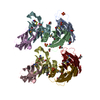
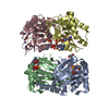
- PDB-1hql: The xenograft antigen in complex with the B4 isolectin of Griffon... -
+ Open data

- Basic information
| Entry | Database: PDB / ID: 1hql | |||||||||
|---|---|---|---|---|---|---|---|---|---|---|
| Title | The xenograft antigen in complex with the B4 isolectin of Griffonia simplicifolia lectin-1 | |||||||||
 Components Components | LECTIN | |||||||||
 Keywords Keywords | SUGAR BINDING PROTEIN / Griffonia simplicifolia / lectin / xenograft antigen | |||||||||
| Function / homology |  Function and homology information Function and homology information | |||||||||
| Biological species |  Griffonia simplicifolia (plant) Griffonia simplicifolia (plant) | |||||||||
| Method | X-RAY DIFFRACTION / Resolution: 2.2 Å | |||||||||
 Authors Authors | Tempel, W. / Lipscomb, L.A. / Rose, J.P. / Woods, R.J. | |||||||||
 Citation Citation | Journal: J.Biol.Chem. / Year: 2002 Title: The xenograft antigen bound to Griffonia simplicifolia lectin 1-B(4). X-ray crystal structure of the complex and molecular dynamics characterization of the binding site. Authors: Tempel, W. / Tschampel, S. / Woods, R.J. | |||||||||
| History |
|
- Structure visualization
| Structure viewer | Molecule: MolmilJmol/JSmol |
|---|
- Downloads & links
-Download
| PDBx/mmCIF format | 1hql.cif.gz | 106.1 KB | Display | PDBx/mmCIF format |
|---|---|---|---|---|
| PDB format | pdb1hql.ent.gz | 84.8 KB | Display | PDB format |
| PDBx/mmJSON format | 1hql.json.gz | Tree view | PDBx/mmJSON format | |
| Others |  Other downloads Other downloads |
-Validation report
| Arichive directory | https://data.pdbj.org/pub/pdb/validation_reports/hq/1hqlftp://data.pdbj.org/pub/pdb/validation_reports/hq/1hql | HTTPS FTP |
|---|
-Related structure data
| Related structure data | |
|---|---|
| Similar structure data |


-Links
 PDBj
PDBj

- Assembly
| Deposited unit | 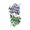
| ||||||||
|---|---|---|---|---|---|---|---|---|---|
| 1 | 
| ||||||||
| Unit cell |
| ||||||||
| Components on special symmetry positions |
| ||||||||
| Details | The full tetramer is generated by applying the following operation to the content of the asymmetric unit: 1-x,-y,z |
-Components
-Protein , 1 types, 2 molecules AB
| #1: Protein | Mass: 28313.207 Da / Num. of mol.: 2 / Source method: isolated from a natural source / Source: (natural) Griffonia simplicifolia (plant) / Organ: SEED / References: UniProt: Q8W1R6 |
|---|
-Sugars , 2 types, 4 molecules
| #2: Polysaccharide | / Mass: 356.323 Da / Num. of mol.: 2 Source method: isolated from a genetically manipulated source #3: Polysaccharide | / Mass: 424.401 Da / Num. of mol.: 2Source method: isolated from a genetically manipulated source |
|---|
-Non-polymers , 3 types, 115 molecules 


| #4: Chemical |  Mass: 54.938 Da / Num. of mol.: 2 / Source method: obtained synthetically / Formula: Mn Mass: 54.938 Da / Num. of mol.: 2 / Source method: obtained synthetically / Formula: Mn#5: Chemical |  Mass: 40.078 Da / Num. of mol.: 2 / Source method: obtained synthetically / Formula: Ca Mass: 40.078 Da / Num. of mol.: 2 / Source method: obtained synthetically / Formula: Ca#6: Water | ChemComp-HOH / | WaterMass: 18.015 Da / Num. of mol.: 111 / Source method: isolated from a natural source / Formula: H2O |
|---|
-Experimental details
-Experiment
| Experiment | Method: X-RAY DIFFRACTION / Number of used crystals: 2 |
|---|
- Sample preparation
| Crystal | Density Matthews: 1.94 Å3/Da / Density % sol: 36.62 % | ||||||||||||||||||||||||||||||||||||||||||
|---|---|---|---|---|---|---|---|---|---|---|---|---|---|---|---|---|---|---|---|---|---|---|---|---|---|---|---|---|---|---|---|---|---|---|---|---|---|---|---|---|---|---|---|
| Crystal grow | Temperature: 293 K / Method: vapor diffusion, hanging drop / pH: 7.5 Details: 5mg/mL protein, approx. 5 equ. sugar, 11% w/v PEG 4000, 8% v/v MPD, 6% v/v DMSO, 0.1M HEPES pH7.5, VAPOR DIFFUSION, HANGING DROP, temperature 293K | ||||||||||||||||||||||||||||||||||||||||||
| Crystal grow | *PLUS Details: Tempel, W., (2001) Acta Crystallogr., D57, 1639. | ||||||||||||||||||||||||||||||||||||||||||
| Components of the solutions | *PLUS
|
-Data collection
| Diffraction |
| |||||||||||||||
|---|---|---|---|---|---|---|---|---|---|---|---|---|---|---|---|---|
| Diffraction source | Source: ROTATING ANODE / Type: RIGAKU RU200 / Wavelength: 1.5418 Å | |||||||||||||||
| Detector |
| |||||||||||||||
| Radiation |
| |||||||||||||||
| Radiation wavelength | Wavelength: 1.5418 Å / Relative weight: 1 | |||||||||||||||
| Reflection | Resolution: 2.2→20 Å / Num. obs: 22043 / % possible obs: 96.7 % / Observed criterion σ(I): -3 / Biso Wilson estimate: 42.6 Å2 / Rmerge(I) obs: 0.142 / Net I/σ(I): 16 | |||||||||||||||
| Reflection shell | Resolution: 2.2→2.3 Å / Rmerge(I) obs: 0.416 / Mean I/σ(I) obs: 4.6 / Num. unique all: 2325 / % possible all: 83.8 | |||||||||||||||
| Reflection | *PLUS Highest resolution: 2.2 Å / Lowest resolution: 20 Å / Num. all: 23055 | |||||||||||||||
| Reflection shell | *PLUS % possible obs: 83.8 % |
- Processing
| Software |
| ||||||||||||||||||||||||||||||||||||||||||||
|---|---|---|---|---|---|---|---|---|---|---|---|---|---|---|---|---|---|---|---|---|---|---|---|---|---|---|---|---|---|---|---|---|---|---|---|---|---|---|---|---|---|---|---|---|---|
| Refinement | Resolution: 2.2→19.93 Å / Rfactor Rfree error: 0.007 / Isotropic thermal model: RESTRAINED / Cross valid method: THROUGHOUT / σ(F): 0 Details: Occupancy 0.00 signifies atoms with missing or uninterpretable density and is not the result of occupancy refinement. The first 3 N-terminal and last 18 C-terminal residues are missing in the electron density.
| ||||||||||||||||||||||||||||||||||||||||||||
| Solvent computation | Solvent model: flat model / Bsol: 115.319 Å2 / ksol: 0.47181 e/Å3 | ||||||||||||||||||||||||||||||||||||||||||||
| Displacement parameters | Biso mean: 38.9 Å2
| ||||||||||||||||||||||||||||||||||||||||||||
| Refine analyze |
| ||||||||||||||||||||||||||||||||||||||||||||
| Refinement step | Cycle: LAST / Resolution: 2.2→19.93 Å
| ||||||||||||||||||||||||||||||||||||||||||||
| Refine LS restraints |
| ||||||||||||||||||||||||||||||||||||||||||||
| LS refinement shell | Resolution: 2.2→2.34 Å / Rfactor Rfree error: 0.02 / Total num. of bins used: 6
| ||||||||||||||||||||||||||||||||||||||||||||
| Xplor file |
| ||||||||||||||||||||||||||||||||||||||||||||
| Software | *PLUS Name: CNS / Version: 1 / Classification: refinement | ||||||||||||||||||||||||||||||||||||||||||||
| Refinement | *PLUS σ(F): 0 / % reflection Rfree: 6.8 % / Rfactor obs: 0.233 | ||||||||||||||||||||||||||||||||||||||||||||
| Solvent computation | *PLUS | ||||||||||||||||||||||||||||||||||||||||||||
| Displacement parameters | *PLUS Biso mean: 38.9 Å2 | ||||||||||||||||||||||||||||||||||||||||||||
| Refine LS restraints | *PLUS
| ||||||||||||||||||||||||||||||||||||||||||||
| LS refinement shell | *PLUS Rfactor Rfree: 0.286 / Rfactor Rwork: 0.254 |
