 Movie
Movie Controller


+ Open data

- Basic information
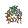
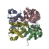
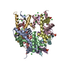
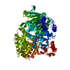
| Entry | Database: PDB / ID: 1hq3 | ||||||
|---|---|---|---|---|---|---|---|
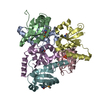
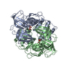
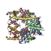
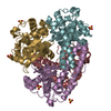
| Title | CRYSTAL STRUCTURE OF THE HISTONE-CORE-OCTAMER IN KCL/PHOSPHATE | ||||||
 Components Components |
| ||||||
 Keywords Keywords |  DNA BINDING PROTEIN / histone-core octamer / nucleosome / chromatin / structural elements / histone-tail guide helices / tether structures / Cl and Phosphate ions / waters DNA BINDING PROTEIN / histone-core octamer / nucleosome / chromatin / structural elements / histone-tail guide helices / tether structures / Cl and Phosphate ions / waters | ||||||
| Function / homology |  Function and homology information Function and homology informationPKMTs methylate histone lysines / HDMs demethylate histones / RMTs methylate histone arginines / SUMOylation of chromatin organization proteins / Condensation of Prophase Chromosomes / Nonhomologous End-Joining (NHEJ) / G2/M DNA damage checkpoint / Metalloprotease DUBs / Processing of DNA double-strand break ends / Recruitment and ATM-mediated phosphorylation of repair and signaling proteins at DNA double strand breaks ...PKMTs methylate histone lysines / HDMs demethylate histones / RMTs methylate histone arginines / SUMOylation of chromatin organization proteins / Condensation of Prophase Chromosomes / Nonhomologous End-Joining (NHEJ) / G2/M DNA damage checkpoint / Metalloprotease DUBs / Processing of DNA double-strand break ends / Recruitment and ATM-mediated phosphorylation of repair and signaling proteins at DNA double strand breaks / Interleukin-7 signaling / Chromatin modifying enzymes / HATs acetylate histones / Ub-specific processing proteases / PRC2 methylates histones and DNA / Oxidative Stress Induced Senescence / B-WICH complex positively regulates rRNA expression / Transcriptional regulation by small RNAs / Assembly of the ORC complex at the origin of replication / RNA Polymerase I Promoter Opening / RNA Polymerase I Promoter Escape / RUNX1 regulates genes involved in megakaryocyte differentiation and platelet function / Estrogen-dependent gene expression / Deposition of new CENPA-containing nucleosomes at the centromere / Factors involved in megakaryocyte development and platelet production / nucleosome assembly / structural constituent of chromatin / nucleosome / protein heterodimerization activity / protein-containing complex binding / DNA binding / nucleoplasm / nucleusSimilarity search - Function | ||||||
| Biological species |  Gallus gallus (chicken) Gallus gallus (chicken) | ||||||
| Method | X-RAY DIFFRACTION / SYNCHROTRON / MOLECULAR REPLACEMENT / Resolution: 2.15 Å | ||||||
 Authors Authors | Chantalat, L. / Nicholson, J.M. / Lambert, S.J. / Reid, A.J. / Donovan, M.J. / Reynolds, C.D. / Wood, C.M. / Baldwin, J.P. | ||||||
 Citation Citation | Journal: Acta Crystallogr.,Sect.D / Year: 2003 Title: Structure of the histone-core octamer in KCl/phosphate crystals at 2.15 A resolution. Authors: Chantalat, L. / Nicholson, J.M. / Lambert, S.J. / Reid, A.J. / Donovan, M.J. / Reynolds, C.D. / Wood, C.M. / Baldwin, J.P. | ||||||
| History |
|
- Structure visualization
| Structure viewer | Molecule: MolmilJmol/JSmol |
|---|
- Downloads & links
-Download
| PDBx/mmCIF format | 1hq3.cif.gz | 174.7 KB | Display | PDBx/mmCIF format |
|---|---|---|---|---|
| PDB format | pdb1hq3.ent.gz | 136.5 KB | Display | PDB format |
| PDBx/mmJSON format | 1hq3.json.gz | Tree view | PDBx/mmJSON format | |
| Others |  Other downloads Other downloads |
-Validation report
| Arichive directory | https://data.pdbj.org/pub/pdb/validation_reports/hq/1hq3ftp://data.pdbj.org/pub/pdb/validation_reports/hq/1hq3 | HTTPS FTP |
|---|
-Related structure data
| Related structure data | 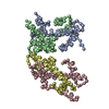 1hioS S: Starting model for refinement |
|---|---|
| Similar structure data |


-Links
 PDBj
PDBj

- Assembly
| Deposited unit | 
| ||||||||
|---|---|---|---|---|---|---|---|---|---|
| 1 |
| ||||||||
| Unit cell |
|
-Components
-Protein , 4 types, 8 molecules AEBFCGDH
| #1: Protein | Mass: 13969.363 Da / Num. of mol.: 2 / Source method: isolated from a natural source / Details: EXTRACTED AS PART OF OCTAMER IN KCL/PHOSPHATE / Source: (natural) Gallus gallus (chicken) / Organelle: CHICK-ERYTHROCYTE NUCLEI / References: UniProt: P02263#2: Protein | Mass: 13953.251 Da / Num. of mol.: 2 / Source method: isolated from a natural source / Details: EXTRACTED AS PART OF OCTAMER IN KCL/PHOSPHATE / Source: (natural) Gallus gallus (chicken) / Organelle: CHICK-ERYTHROCYTE NUCLEI / References: UniProt: P02279, UniProt: P0C1H5*PLUS#3: Protein | Mass: 15421.101 Da / Num. of mol.: 2 / Source method: isolated from a natural source / Details: EXTRACTED AS PART OF OCTAMER IN KCL/PHOSPHATE / Source: (natural) Gallus gallus (chicken) / Organelle: CHICK-ERYTHROCYTE NUCLEI / References: UniProt: P84229#4: Protein | Mass: 11394.426 Da / Num. of mol.: 2 / Source method: isolated from a natural source / Details: EXTRACTED AS PART OF OCTAMER IN KCL/PHOSPHATE / Source: (natural) Gallus gallus (chicken) / Organelle: CHICK-ERYTHROCYTE NUCLEI / References: UniProt: P62801 |
|---|
-Non-polymers , 3 types, 464 molecules 


| #5: Chemical | ChemComp-PO4 / Phosphate Mass: 94.971 Da / Num. of mol.: 5 / Source method: obtained synthetically / Formula: PO4 Mass: 94.971 Da / Num. of mol.: 5 / Source method: obtained synthetically / Formula: PO4#6: Chemical | ChemComp-CL / Chloride Mass: 35.453 Da / Num. of mol.: 22 / Source method: obtained synthetically / Formula: Cl Mass: 35.453 Da / Num. of mol.: 22 / Source method: obtained synthetically / Formula: Cl#7: Water | ChemComp-HOH / | WaterMass: 18.015 Da / Num. of mol.: 437 / Source method: isolated from a natural source / Formula: H2O |
|---|
-Experimental details
-Experiment
| Experiment | Method: X-RAY DIFFRACTION / Number of used crystals: 2 |
|---|
- Sample preparation
| Crystal | Density Matthews: 3.45 Å3/Da / Density % sol: 64.4 % | ||||||||||||||||||||||||||||||||||||||||||
|---|---|---|---|---|---|---|---|---|---|---|---|---|---|---|---|---|---|---|---|---|---|---|---|---|---|---|---|---|---|---|---|---|---|---|---|---|---|---|---|---|---|---|---|
| Crystal grow | Method: microdialysis / pH: 6.7 Details: 2.0M KCL, 1.35M phosphate, pH 6.6, pH 6.7, MICRODIALYSIS | ||||||||||||||||||||||||||||||||||||||||||
| Crystal grow | *PLUS Temperature: 277 K / pH: 6.9 | ||||||||||||||||||||||||||||||||||||||||||
| Components of the solutions | *PLUS
|
-Data collection
| Diffraction |
| ||||||||||||||||||
|---|---|---|---|---|---|---|---|---|---|---|---|---|---|---|---|---|---|---|---|
| Diffraction source |
| ||||||||||||||||||
| Detector |
| ||||||||||||||||||
| Radiation |
| ||||||||||||||||||
| Radiation wavelength | Wavelength: 1.488 Å / Relative weight: 1 | ||||||||||||||||||
| Reflection | Resolution: 2.15→20 Å / Num. all: 74806 / Num. obs: 74806 / % possible obs: 97.4 % / Redundancy: 2.26 % / Rmerge(I) obs: 0.071 | ||||||||||||||||||
| Reflection shell | Resolution: 2.15→2.25 Å / Redundancy: 2.16 % / Rmerge(I) obs: 0.331 / % possible all: 97.1 | ||||||||||||||||||
| Reflection | *PLUS Num. measured all: 169058 | ||||||||||||||||||
| Reflection shell | *PLUS % possible obs: 97.1 % |

- Processing
| Software |
| ||||||||||||||||||||||||||||||||||||||||
|---|---|---|---|---|---|---|---|---|---|---|---|---|---|---|---|---|---|---|---|---|---|---|---|---|---|---|---|---|---|---|---|---|---|---|---|---|---|---|---|---|---|
| Refinement | Method to determine structure: MOLECULAR REPLACEMENT Starting model: histone octamer PDB entry 1HIO Resolution: 2.15→20 Å / Rfactor Rfree error: 0.004 / Isotropic thermal model: restrained / Cross valid method: THROUGHOUT / σ(F): 0 / σ(I): 0
| ||||||||||||||||||||||||||||||||||||||||
| Solvent computation | Solvent model: flat model / Bsol: 61.1947 Å2 / ksol: 0.371738 e/Å3 | ||||||||||||||||||||||||||||||||||||||||
| Displacement parameters | Biso mean: 40.8 Å2 | ||||||||||||||||||||||||||||||||||||||||
| Refine analyze |
| ||||||||||||||||||||||||||||||||||||||||
| Refinement step | Cycle: LAST / Resolution: 2.15→20 Å
| ||||||||||||||||||||||||||||||||||||||||
| Refine LS restraints |
| ||||||||||||||||||||||||||||||||||||||||
| LS refinement shell | Resolution: 2.15→2.28 Å / Total num. of bins used: 6 / % reflection obs: 97.1 % | ||||||||||||||||||||||||||||||||||||||||
| Xplor file |
| ||||||||||||||||||||||||||||||||||||||||
| Software | *PLUS Name: CNS / Classification: refinement | ||||||||||||||||||||||||||||||||||||||||
| Refinement | *PLUS σ(F): 0 / % reflection Rfree: 6 % / Rfactor obs: 0.214 | ||||||||||||||||||||||||||||||||||||||||
| Solvent computation | *PLUS | ||||||||||||||||||||||||||||||||||||||||
| Displacement parameters | *PLUS Biso mean: 40.8 Å2 | ||||||||||||||||||||||||||||||||||||||||
| Refine LS restraints | *PLUS
|
