 Movie
Movie Controller

[English] 日本語
 Yorodumi
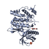
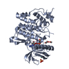
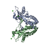
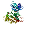
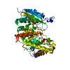
- PDB-1how: THE X-RAY CRYSTAL STRUCTURE OF SKY1P, AN SR PROTEIN KINASE IN YEAST -
+ Open data

- Basic information
| Entry | Database: PDB / ID: 1how | ||||||
|---|---|---|---|---|---|---|---|
| Title | THE X-RAY CRYSTAL STRUCTURE OF SKY1P, AN SR PROTEIN KINASE IN YEAST | ||||||
 Components Components | SERINE/THREONINE-PROTEIN KINASE YMR216C | ||||||
 Keywords Keywords |  TRANSFERASE / KINASE TRANSFERASE / KINASE | ||||||
| Function / homology |  Function and homology information Function and homology informationintracellular monoatomic cation homeostasis / intracellular monoatomic ion homeostasis / regulation of mRNA processing / mRNA splice site recognition / stress granule disassembly / regulation of cell size / spliceosomal complex assembly / cytoplasmic stress granule / positive regulation of protein import into nucleus / non-specific serine/threonine protein kinase ...intracellular monoatomic cation homeostasis / intracellular monoatomic ion homeostasis / regulation of mRNA processing / mRNA splice site recognition / stress granule disassembly / regulation of cell size / spliceosomal complex assembly / cytoplasmic stress granule / positive regulation of protein import into nucleus / non-specific serine/threonine protein kinase / protein kinase activity / response to xenobiotic stimulus / phosphorylation / protein serine kinase activity / protein serine/threonine kinase activity / ATP binding / nucleus / cytoplasmSimilarity search - Function | ||||||
| Biological species |  Saccharomyces cerevisiae (brewer's yeast) Saccharomyces cerevisiae (brewer's yeast) | ||||||
| Method | X-RAY DIFFRACTION / MIR / Resolution: 2.1 Å | ||||||
 Authors Authors | Nolen, B.J. / Yun, C.Y. / Wong, C.F. / McCammon, J.A. / Fu, X.-D. / Ghosh, G. | ||||||
 Citation Citation | Journal: Nat.Struct.Biol. / Year: 2001 Title: The structure of Sky1p reveals a novel mechanism for constitutive activity. Authors: Nolen, B. / Yun, C.Y. / Wong, C.F. / McCammon, J.A. / Fu, X.D. / Ghosh, G. | ||||||
| History |
|
- Structure visualization
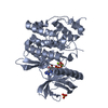
| Structure viewer | Molecule: MolmilJmol/JSmol |
|---|
- Downloads & links
-Download
| PDBx/mmCIF format | 1how.cif.gz | 87.3 KB | Display | PDBx/mmCIF format |
|---|---|---|---|---|
| PDB format | pdb1how.ent.gz | 65.6 KB | Display | PDB format |
| PDBx/mmJSON format | 1how.json.gz | Tree view | PDBx/mmJSON format | |
| Others |  Other downloads Other downloads |
-Validation report
| Arichive directory | https://data.pdbj.org/pub/pdb/validation_reports/ho/1howftp://data.pdbj.org/pub/pdb/validation_reports/ho/1how | HTTPS FTP |
|---|
-Related structure data
| Similar structure data |
|---|


-Links
 PDBj
PDBj- Assembly
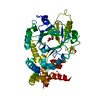
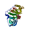
| Deposited unit | 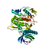
| ||||||||
|---|---|---|---|---|---|---|---|---|---|
| 1 |
| ||||||||
| Unit cell |
|
-Components
| #1: Protein | Mass: 43009.066 Da / Num. of mol.: 1 / Fragment: SKY1PDELTANS Mutation: 137 A.A. TRUNCATED FROM N-TERMINUS, RESIDUES 305-542 REMOVED AND REPLACED WITH VDSQK Source method: isolated from a genetically manipulated source Source: (gene. exp.) Saccharomyces cerevisiae (brewer's yeast)Gene: YMR216C / Plasmid: PET15B / Production host:  Escherichia coli (E. coli) / Strain (production host): BL21(DE3)PLYSS Escherichia coli (E. coli) / Strain (production host): BL21(DE3)PLYSSReferences: UniProt: Q03656, Transferases; Transferring phosphorus-containing groups; Phosphotransferases with an alcohol group as acceptor | ||||
|---|---|---|---|---|---|
| #2: Chemical | ChemComp-SO4 / Sulfate  Mass: 96.063 Da / Num. of mol.: 6 / Source method: obtained synthetically / Formula: SO4 Mass: 96.063 Da / Num. of mol.: 6 / Source method: obtained synthetically / Formula: SO4#3: Chemical | Ethylene glycol  Mass: 62.068 Da / Num. of mol.: 2 / Source method: obtained synthetically / Formula: C2H6O2 Mass: 62.068 Da / Num. of mol.: 2 / Source method: obtained synthetically / Formula: C2H6O2#4: Water | ChemComp-HOH / | Water Mass: 18.015 Da / Num. of mol.: 204 / Source method: isolated from a natural source / Formula: H2O Mass: 18.015 Da / Num. of mol.: 204 / Source method: isolated from a natural source / Formula: H2O |
-Experimental details
-Experiment
| Experiment | Method: X-RAY DIFFRACTION / Number of used crystals: 1 |
|---|
- Sample preparation
| Crystal | Density Matthews: 2.56 Å3/Da / Density % sol: 51.96 % | ||||||||||||||||||||||||||||||||||||||||||
|---|---|---|---|---|---|---|---|---|---|---|---|---|---|---|---|---|---|---|---|---|---|---|---|---|---|---|---|---|---|---|---|---|---|---|---|---|---|---|---|---|---|---|---|
| Crystal grow | Temperature: 291 K / Method: vapor diffusion, hanging drop / pH: 4.5 Details: Ammonium Sulfate, Ethylene Glycol, Sodium Acetate, pH 4.5, VAPOR DIFFUSION, HANGING DROP, temperature 291.0K | ||||||||||||||||||||||||||||||||||||||||||
| Crystal | *PLUS Density % sol: 52 % | ||||||||||||||||||||||||||||||||||||||||||
| Crystal grow | *PLUS | ||||||||||||||||||||||||||||||||||||||||||
| Components of the solutions | *PLUS
|
-Data collection
| Diffraction | Mean temperature: 105 K |
|---|---|
| Diffraction source | Source: ROTATING ANODE / Type: RIGAKU / Wavelength: 1.54 Å |
| Detector | Type: MARRESEARCH / Detector: IMAGE PLATE / Date: Apr 8, 1999 / Details: mirrors |
| Radiation | Monochromator: MIRRORS / Protocol: SINGLE WAVELENGTH / Monochromatic (M) / Laue (L): M / Scattering type: x-ray |
| Radiation wavelength | Wavelength: 1.54 Å / Relative weight: 1 |
| Reflection | Resolution: 2.1→20 Å / Num. all: 29021 / Num. obs: 24675 / % possible obs: 92.4 % / Observed criterion σ(F): 2 / Observed criterion σ(I): 1 / Redundancy: 8.1 % / Rmerge(I) obs: 0.046 / Net I/σ(I): 30.6 |
| Reflection shell | Resolution: 2.1→2.19 Å / Rmerge(I) obs: 0.533 / Mean I/σ(I) obs: 3.3 / % possible all: 91.8 |
| Reflection | *PLUS Num. measured all: 235953 |
| Reflection shell | *PLUS % possible obs: 91.8 % |
- Processing
| Software |
| ||||||||||||||||||||
|---|---|---|---|---|---|---|---|---|---|---|---|---|---|---|---|---|---|---|---|---|---|
| Refinement | Method to determine structure: MIR / Resolution: 2.1→20 Å / σ(F): 2 / σ(I): 1 / Stereochemistry target values: Engh & Huber Details: RESIDUES MENTIONED IN REMARK 470 WERE MODELED AS ALA DUE TO POOR ELECTRON DENSITY
| ||||||||||||||||||||
| Refinement step | Cycle: LAST / Resolution: 2.1→20 Å
| ||||||||||||||||||||
| Refine LS restraints |
| ||||||||||||||||||||
| Software | *PLUS Name: CNS / Classification: refinement | ||||||||||||||||||||
| Refinement | *PLUS Highest resolution: 2.1 Å / Lowest resolution: 20 Å / σ(F): 2 / Rfactor obs: 0.204 | ||||||||||||||||||||
| Solvent computation | *PLUS | ||||||||||||||||||||
| Displacement parameters | *PLUS |
