 Movie
Movie Controller

[English] 日本語
 Yorodumi
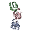
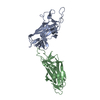
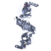
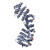
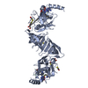
- PDB-1ho8: CRYSTAL STRUCTURE OF THE REGULATORY SUBUNIT H OF THE V-TYPE ATPAS... -
+ Open data

- Basic information
| Entry | Database: PDB / ID: 1ho8 | ||||||
|---|---|---|---|---|---|---|---|
| Title | CRYSTAL STRUCTURE OF THE REGULATORY SUBUNIT H OF THE V-TYPE ATPASE OF SACCHAROMYCES CEREVISIAE | ||||||
 Components Components | VACUOLAR ATP SYNTHASE SUBUNIT H | ||||||
 Keywords Keywords |  HYDROLASE / HEAT repeat HYDROLASE / HEAT repeat | ||||||
| Function / homology |  Function and homology information Function and homology informationInsulin receptor recycling / Transferrin endocytosis and recycling / ROS and RNS production in phagocytes / Amino acids regulate mTORC1 / Golgi lumen acidification / vacuolar proton-transporting V-type ATPase, V1 domain / endosomal lumen acidification / proton-transporting V-type ATPase complex / vacuolar proton-transporting V-type ATPase complex / vacuolar acidification ...Insulin receptor recycling / Transferrin endocytosis and recycling / ROS and RNS production in phagocytes / Amino acids regulate mTORC1 / Golgi lumen acidification / vacuolar proton-transporting V-type ATPase, V1 domain / endosomal lumen acidification / proton-transporting V-type ATPase complex / vacuolar proton-transporting V-type ATPase complex / vacuolar acidification / fungal-type vacuole membrane / proton-transporting ATPase activity, rotational mechanism / proton transmembrane transport / Golgi membraneSimilarity search - Function | ||||||
| Biological species |  Saccharomyces cerevisiae (brewer's yeast) Saccharomyces cerevisiae (brewer's yeast) | ||||||
| Method | X-RAY DIFFRACTION / SYNCHROTRON / SIR, MAD / Resolution: 2.95 Å | ||||||
 Authors Authors | Sagermann, M. / Stevens, T.H. / Matthews, B.W. | ||||||
 Citation Citation | Journal: Proc.Natl.Acad.Sci.USA / Year: 2001 Title: Crystal structure of the regulatory subunit H of the V-type ATPase of Saccharomyces cerevisiae. Authors: Sagermann, M. / Stevens, T.H. / Matthews, B.W. #1: Journal: Acta Crystallogr.,Sect.D / Year: 2000Title: Cloning, expression and crystallization of VMA13p, an essential subunit of the vacuolar H+-ATPase of Saccharomyces cerevisiae. Authors: Sagermann, M. / Matthews, B.W. #2: Journal: J.Biol.Chem. / Year: 1993Title: VMA13 encodes a 54-kDa vacuolar H(+)-ATPase subunit required for activity but not assembly of the enzyme complex in Saccharomyces cerevisiae. Authors: Ho, M.N. / Hirata, R. / Umemoto, N. / Ohya, Y. / Takatsuki, A. / Stevens, T.H. / Anraku, Y. | ||||||
| History |
|
- Structure visualization
| Structure viewer | Molecule: MolmilJmol/JSmol |
|---|
- Downloads & links
-Download
| PDBx/mmCIF format | 1ho8.cif.gz | 102.9 KB | Display | PDBx/mmCIF format |
|---|---|---|---|---|
| PDB format | pdb1ho8.ent.gz | 78.4 KB | Display | PDB format |
| PDBx/mmJSON format | 1ho8.json.gz | Tree view | PDBx/mmJSON format | |
| Others |  Other downloads Other downloads |
-Validation report
| Arichive directory | https://data.pdbj.org/pub/pdb/validation_reports/ho/1ho8ftp://data.pdbj.org/pub/pdb/validation_reports/ho/1ho8 | HTTPS FTP |
|---|
-Related structure data
| Similar structure data |
|---|


-Links
 PDBj
PDBj

- Assembly
| Deposited unit | 
| ||||||||
|---|---|---|---|---|---|---|---|---|---|
| 1 |
| ||||||||
| Unit cell |
|
-Components
| #1: Protein | Mass: 54626.738 Da / Num. of mol.: 1 Source method: isolated from a genetically manipulated source Source: (gene. exp.) Saccharomyces cerevisiae (brewer's yeast)Plasmid: PGEX4T3 / Species (production host): Escherichia coli / Production host:  Escherichia coli BL21(DE3) (bacteria) / Strain (production host): BL21(DE3) / References: UniProt: P41807, EC: 3.6.1.34 Escherichia coli BL21(DE3) (bacteria) / Strain (production host): BL21(DE3) / References: UniProt: P41807, EC: 3.6.1.34 |
|---|---|
| #2: Chemical | ChemComp-SO4 / Sulfate  Mass: 96.063 Da / Num. of mol.: 1 / Source method: obtained synthetically / Formula: SO4 Mass: 96.063 Da / Num. of mol.: 1 / Source method: obtained synthetically / Formula: SO4 |
| #3: Water | ChemComp-HOH / Water Mass: 18.015 Da / Num. of mol.: 25 / Source method: isolated from a natural source / Formula: H2O Mass: 18.015 Da / Num. of mol.: 25 / Source method: isolated from a natural source / Formula: H2O |
-Experimental details
-Experiment
| Experiment | Method: X-RAY DIFFRACTION / Number of used crystals: 1 |
|---|
- Sample preparation
| Crystal | Density Matthews: 4.36 Å3/Da / Density % sol: 71.79 % | ||||||||||||||||||||||||||||||||||||||||||||||||||||||
|---|---|---|---|---|---|---|---|---|---|---|---|---|---|---|---|---|---|---|---|---|---|---|---|---|---|---|---|---|---|---|---|---|---|---|---|---|---|---|---|---|---|---|---|---|---|---|---|---|---|---|---|---|---|---|---|
| Crystal grow | Temperature: 298 K / Method: vapor diffusion, hanging drop / pH: 7.5 Details: Ammonium sulfate, Dithiothreitol, pH 7.5, VAPOR DIFFUSION, HANGING DROP, temperature 298K | ||||||||||||||||||||||||||||||||||||||||||||||||||||||
| Crystal grow | *PLUS Details: Sagermann, M., (2000) Acta Crystallogr., Sect.D, 56, 475. | ||||||||||||||||||||||||||||||||||||||||||||||||||||||
| Components of the solutions | *PLUS
|
-Data collection
| Diffraction | Ambient pressure: 101 kPa / Mean temperature: 100 K |
|---|---|
| Diffraction source | Source: SYNCHROTRON / Site: SSRL  / Beamline: BL9-2 / Wavelength: 0.979 Å / Beamline: BL9-2 / Wavelength: 0.979 Å |
| Detector | Type: ADSC QUANTUM 4 / Detector: CCD / Date: Nov 24, 2000 |
| Radiation | Monochromator: Double-Crystal Si 111 crystals / Protocol: MAD / Monochromatic (M) / Laue (L): M / Scattering type: x-ray / Wavelength: 0.979 Å |
| Radiation wavelength | Wavelength: 0.979 Å / Relative weight: 1 |
| Reflection | Resolution: 2.95→71 Å / Num. all: 20090 / Num. obs: 20090 / % possible obs: 99.8 % / Observed criterion σ(F): 0 / Observed criterion σ(I): 0 / Redundancy: 4.3 % / Biso Wilson estimate: 65 Å2 / Rmerge(I) obs: 0.077 / Net I/σ(I): 7.4 |
| Reflection shell | Resolution: 2.95→3.3 Å / Redundancy: 4.3 % / Rmerge(I) obs: 0.454 / Mean I/σ(I) obs: 1.7 / Num. unique all: 5733 / Rsym value: 45.4 / % possible all: 99.9 |
| Reflection | *PLUS Num. measured all: 87426 |
- Processing
| Software |
| |||||||||||||||||||||||||
|---|---|---|---|---|---|---|---|---|---|---|---|---|---|---|---|---|---|---|---|---|---|---|---|---|---|---|
| Refinement | Method to determine structure: SIR, MAD Starting model: vma13 poly alanine Resolution: 2.95→71 Å / Isotropic thermal model: Anisotropic / Cross valid method: THROUGHOUT / σ(F): 0 / σ(I): 0 / Stereochemistry target values: Engh & Huber Details: REFINEMENT AS IMPLEMENTED IN TNT. RESIDUES 54-70 AND 224- 236 WERE NOT MODELED DUE TO DISORDER. RESIDUES 408-415 AND 455-459 WERE PARTIALLY VISIBLE IN THE ELECTRON DENSITY AND REFINED TO HIGH B VALUES
| |||||||||||||||||||||||||
| Displacement parameters |
| |||||||||||||||||||||||||
| Refinement step | Cycle: LAST / Resolution: 2.95→71 Å
| |||||||||||||||||||||||||
| Refine LS restraints |
| |||||||||||||||||||||||||
| LS refinement shell | Resolution: 2.95→71 Å
| |||||||||||||||||||||||||
| Software | *PLUS Name: TNT / Classification: refinement | |||||||||||||||||||||||||
| Refinement | *PLUS σ(F): 0 / % reflection Rfree: 8 % / Rfactor all: 0.223 | |||||||||||||||||||||||||
| Solvent computation | *PLUS | |||||||||||||||||||||||||
| Displacement parameters | *PLUS | |||||||||||||||||||||||||
| Refine LS restraints | *PLUS
|
