[English] 日本語
Yorodumi
- PDB-1hkd: Structure of pea lectin in complex with alpha-methyl-D-glucopyranoside -

+
Open data


ID or keywords:

Loading...

-
Basic information

Entry
Database: PDB / ID: 1hkd
TitleStructure of pea lectin in complex with alpha-methyl-D-glucopyranoside
Components
  • PEA LECTIN ALPHA CHAIN
  • PEA LECTIN BETA CHAIN
KeywordsPLANT LECTIN / CARBOHYDRATE BINDING PROTEIN / CALCIUM / GLYCOPROTEIN
Function / homology
Function and homology information


D-mannose binding / metal ion binding
Similarity search - Function
Legume lectin / Legume lectin, alpha chain, conserved site / Legume lectins alpha-chain signature. / Legume lectins beta-chain signature. / Legume lectin domain / Legume lectin, beta chain, Mn/Ca-binding site / Legume lectin domain / Jelly Rolls - #200 / Concanavalin A-like lectin/glucanase domain superfamily / Jelly Rolls ...Legume lectin / Legume lectin, alpha chain, conserved site / Legume lectins alpha-chain signature. / Legume lectins beta-chain signature. / Legume lectin domain / Legume lectin, beta chain, Mn/Ca-binding site / Legume lectin domain / Jelly Rolls - #200 / Concanavalin A-like lectin/glucanase domain superfamily / Jelly Rolls / Sandwich / Mainly Beta
Similarity search - Domain/homology
methyl alpha-D-glucopyranoside / : / Lectin
Similarity search - Component
Biological speciesPISUM SATIVUM (garden pea)
MethodX-RAY DIFFRACTION / MOLECULAR REPLACEMENT / Resolution: 2.09 Å
AuthorsShevtsov, M.B. / Tsygannik, I.N.
CitationJournal: To be Published
Title: Structure of Pea Lectin in Complex with Alpha-Methyl-D-Glucopyranoside
Authors: Shevtsov, M.B. / Tsygannik, I.N.
History
DepositionMar 6, 2003Deposition site: PDBE / Processing site: PDBE
Revision 1.0Mar 18, 2003Provider: repository / Type: Initial release
Revision 1.1May 8, 2011Group: Version format compliance
Revision 1.2Jul 13, 2011Group: Version format compliance
Revision 1.3Jun 28, 2017Group: Data collection / Category: diffrn_source / Item: _diffrn_source.type
Revision 1.4Jul 29, 2020Group: Data collection / Derived calculations ...Data collection / Derived calculations / Other / Structure summary
Category: chem_comp / entity ...chem_comp / entity / pdbx_chem_comp_identifier / pdbx_database_status / pdbx_entity_nonpoly / pdbx_struct_conn_angle / struct_conn / struct_site / struct_site_gen
Item: _chem_comp.mon_nstd_flag / _chem_comp.name ..._chem_comp.mon_nstd_flag / _chem_comp.name / _entity.pdbx_description / _pdbx_database_status.status_code_sf / _pdbx_entity_nonpoly.name / _pdbx_struct_conn_angle.ptnr1_auth_comp_id / _pdbx_struct_conn_angle.ptnr1_auth_seq_id / _pdbx_struct_conn_angle.ptnr1_label_asym_id / _pdbx_struct_conn_angle.ptnr1_label_atom_id / _pdbx_struct_conn_angle.ptnr1_label_comp_id / _pdbx_struct_conn_angle.ptnr1_label_seq_id / _pdbx_struct_conn_angle.ptnr2_auth_comp_id / _pdbx_struct_conn_angle.ptnr2_auth_seq_id / _pdbx_struct_conn_angle.ptnr2_label_asym_id / _pdbx_struct_conn_angle.ptnr2_label_atom_id / _pdbx_struct_conn_angle.ptnr2_label_comp_id / _pdbx_struct_conn_angle.ptnr3_auth_comp_id / _pdbx_struct_conn_angle.ptnr3_auth_seq_id / _pdbx_struct_conn_angle.ptnr3_label_asym_id / _pdbx_struct_conn_angle.ptnr3_label_atom_id / _pdbx_struct_conn_angle.ptnr3_label_comp_id / _pdbx_struct_conn_angle.ptnr3_label_seq_id / _pdbx_struct_conn_angle.value / _struct_conn.pdbx_dist_value / _struct_conn.ptnr1_auth_comp_id / _struct_conn.ptnr1_auth_seq_id / _struct_conn.ptnr1_label_asym_id / _struct_conn.ptnr1_label_atom_id / _struct_conn.ptnr1_label_comp_id / _struct_conn.ptnr1_label_seq_id / _struct_conn.ptnr2_auth_comp_id / _struct_conn.ptnr2_auth_seq_id / _struct_conn.ptnr2_label_asym_id / _struct_conn.ptnr2_label_atom_id / _struct_conn.ptnr2_label_comp_id / _struct_conn.ptnr2_label_seq_id
Description: Carbohydrate remediation / Provider: repository / Type: Remediation
Revision 1.5Dec 13, 2023Group: Advisory / Data collection ...Advisory / Data collection / Database references / Refinement description / Structure summary
Category: chem_comp / chem_comp_atom ...chem_comp / chem_comp_atom / chem_comp_bond / database_2 / pdbx_initial_refinement_model / pdbx_unobs_or_zero_occ_atoms
Item: _chem_comp.pdbx_synonyms / _database_2.pdbx_DOI / _database_2.pdbx_database_accession
Remark 700 SHEET THE SHEET STRUCTURE OF THIS MOLECULE IS BIFURCATED. IN ORDER TO REPRESENT THIS FEATURE IN ... SHEET THE SHEET STRUCTURE OF THIS MOLECULE IS BIFURCATED. IN ORDER TO REPRESENT THIS FEATURE IN THE SHEET RECORDS BELOW, TWO SHEETS ARE DEFINED.

-
Structure visualization

Structure viewerMolecule:
MolmilJmol/JSmol

Downloads & links

-
Assembly

Deposited unit
A: PEA LECTIN ALPHA CHAIN
B: PEA LECTIN BETA CHAIN
C: PEA LECTIN ALPHA CHAIN
D: PEA LECTIN BETA CHAIN
hetero molecules


Theoretical massNumber of molelcules
Total (without water)51,77310
Polymers51,1944
Non-polymers5786
Water5,098283
1


  • Idetical with deposited unit
  • defined by author&software
TypeNameSymmetry operationNumber
identity operation1_555x,y,z1
MethodPQS
Unit cell
Length a, b, c (Å)50.793, 61.235, 136.005
Angle α, β, γ (deg.)90.00, 90.00, 90.00
Int Tables number19
Space group name H-MP212121
Noncrystallographic symmetry (NCS)NCS oper:
IDCodeMatrix
1given(1), (1), (1)
2given(1), (1), (1)
Details
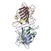
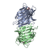
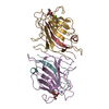
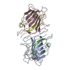
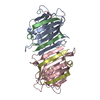
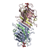
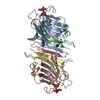
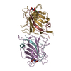
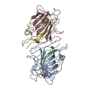
FOR THE HETERO-ASSEMBLY DESCRIBED BY REMARK 350THE PEA LECTIN MOLECULE NORMALLY EXISTS AS A DIMER. THETWO MONOMERS ARE RELATED BY A PSUEDO TWOFOLD AXIS. EACHMONOMER CONSISTS OF TWO SEPARATE POLYPEPTIDE CHAINS, ALPHAAND BETA. THE ALPHA CHAIN CONSISTS OF 181 RESIDUES AND THEBETA CHAIN CONSISTS OF 52 RESIDUES. THE ALPHA AND BETACHAINS OF MONOMER 1 HAVE BEEN ASSIGNED CHAIN IDENTIFIERS*A* AND *B*, RESPECTIVELY, IN THIS ENTRY. THE ALPHA ANDBETA CHAINS OF MONOMER 2 HAVE BEEN ASSIGNED CHAINIDENTIFIERS *C* AND *D*, RESPECTIVELY, IN THIS ENTRY.THISNUMBERING SCHEME IS DIFFERENT FROM THAT IN THE EARLIERPUBLICATIONS.

-
Components

-
Protein , 2 types, 4 molecules ACBD

#1: Protein PEA LECTIN ALPHA CHAIN


Mass: 20001.076 Da / Num. of mol.: 2 / Fragment: RESIDUES 31-211 / Source method: isolated from a natural source
Details: ALPHA-METHYL-D-GLUCOPYRANOSIDE IS PRESENT IN TWO BINDING SITES OF MOLECULE
Source: (natural) PISUM SATIVUM (garden pea) / Organ: SEEDSSeed / References: UniProt: P02867
#2: Protein PEA LECTIN BETA CHAIN


Mass: 5596.086 Da / Num. of mol.: 2 / Fragment: RESIDUES 218-269 / Source method: isolated from a natural source
Details: ALPHA-METHYL-D-GLUCOPYRANOSIDE IS PRESENT IN TWO BINDING SITES OF MOLECULE
Source: (natural) PISUM SATIVUM (garden pea) / Organ: SEEDSSeed / References: UniProt: P02867

-
Sugars , 1 types, 2 molecules

#5: Sugar ChemComp-GYP / methyl alpha-D-glucopyranoside / METHYL-ALPHA-D-GLUCOPYRANOSIDE / ALPHA-METHYL-D-GLUCOPYRANOSIDE / methyl alpha-D-glucoside / methyl D-glucoside / methyl glucoside / Methyl group


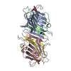
Type: D-saccharide / Mass: 194.182 Da / Num. of mol.: 2
Source method: isolated from a genetically manipulated source
Formula: C7H14O6
IdentifierTypeProgram
DGlcp[1Me]aCONDENSED IUPAC CARBOHYDRATE SYMBOLGMML 1.0
1-methyl-a-D-glucopyranoseCOMMON NAMEGMML 1.0
methyl-a-D-glucopyranosideIUPAC CARBOHYDRATE SYMBOLPDB-CARE 1.0

-
Non-polymers , 3 types, 287 molecules

#3: Chemical ChemComp-CA / CALCIUM ION


Mass: 40.078 Da / Num. of mol.: 2 / Source method: obtained synthetically / Formula: Ca
#4: Chemical ChemComp-MN / MANGANESE (II) ION


Mass: 54.938 Da / Num. of mol.: 2 / Source method: obtained synthetically / Formula: Mn
#6: Water ChemComp-HOH / water / Water


Mass: 18.015 Da / Num. of mol.: 283 / Source method: isolated from a natural source / Formula: H2O

-
Details

Compound detailsD-MANNOSE SPECIFIC LECTIN. TETRAMER OF TWO ALPHA AND TWO BETA CHAINS. BINDS ONE MANGANESE (OR OTHER ...D-MANNOSE SPECIFIC LECTIN. TETRAMER OF TWO ALPHA AND TWO BETA CHAINS. BINDS ONE MANGANESE (OR OTHER TRANSITION METAL) ION AND ONE CALCIUM ION. THE METAL IONS ARE ESSENTIAL FOR THE SACCHARIDE-BINDING AND CELL-AGGLUTINATING ACTIVITIES. SIMILARITY: BELONGS TO THE LEGUMINOUS LECTIN FAMILY.

-
Experimental details

-
Experiment

ExperimentMethod: X-RAY DIFFRACTION / Number of used crystals: 1

-
Sample preparation

CrystalDensity Matthews: 1.94 Å3/Da / Density % sol: 36.18 %
Crystal growpH: 7 / Details: 1.7 M AMMONIUM SULFATE, 10% (V/V) ETHANOL, pH 7.00

-
Data collection

DiffractionMean temperature: 293 K
Diffraction sourceSource: ROTATING ANODE / Type: ELLIOTT GX-6 / Wavelength: 1.5418
DetectorType: MARRESEARCH / Detector: IMAGE PLATE / Date: Jun 15, 2002 / Details: MIRRORS
RadiationMonochromator: NI FILTER / Protocol: SINGLE WAVELENGTH / Monochromatic (M) / Laue (L): M / Scattering type: x-ray
Radiation wavelengthWavelength: 1.5418 Å / Relative weight: 1
ReflectionResolution: 2→30 Å / Num. obs: 30005 / % possible obs: 95.4 % / Redundancy: 2.8 % / Biso Wilson estimate: 28.5 Å2 / Rmerge(I) obs: 0.058 / Net I/σ(I): 17.9
Reflection shellResolution: 2→2.03 Å / Redundancy: 2.19 % / Rmerge(I) obs: 0.42 / Mean I/σ(I) obs: 1.92 / % possible all: 83.5

-
Processing

Software
NameVersionClassification
REFMAC5.1.24refinement
DENZOdata reduction
SCALEPACKdata scaling
MOLREPphasing
RefinementMethod to determine structure: MOLECULAR REPLACEMENT
Starting model: PDB ENTRY 2LTN
Resolution: 2.09→27.95 Å / Cor.coef. Fo:Fc: 0.956 / Cor.coef. Fo:Fc free: 0.937 / SU B: 4.7 / SU ML: 0.124 / Cross valid method: THROUGHOUT / ESU R: 0.243 / ESU R Free: 0.182 / Stereochemistry target values: MAXIMUM LIKELIHOOD
RfactorNum. reflection% reflectionSelection details
Rfree0.209 1181 4.8 %RANDOM
Rwork0.163 ---
obs0.165 23508 98.3 %-
Solvent computationIon probe radii: 0.8 Å / Shrinkage radii: 0.8 Å / VDW probe radii: 1.4 Å / Solvent model: BABINET MODEL WITH MASK
Displacement parametersBiso mean: 22.77 Å2
Baniso -1Baniso -2Baniso -3
1-0.46 Å20 Å20 Å2
2---0.09 Å20 Å2
3----0.36 Å2
Refinement stepCycle: LAST / Resolution: 2.09→27.95 Å
ProteinNucleic acidLigandSolventTotal
Num. atoms3576 0 30 283 3889
Refine LS restraints
Refine-IDTypeDev idealDev ideal targetNumber
X-RAY DIFFRACTIONr_bond_refined_d0.0070.0213687
X-RAY DIFFRACTIONr_bond_other_d
X-RAY DIFFRACTIONr_angle_refined_deg1.3051.9195036
X-RAY DIFFRACTIONr_angle_other_deg
X-RAY DIFFRACTIONr_dihedral_angle_1_deg2.9365454
X-RAY DIFFRACTIONr_dihedral_angle_2_deg
X-RAY DIFFRACTIONr_dihedral_angle_3_deg
X-RAY DIFFRACTIONr_dihedral_angle_4_deg
X-RAY DIFFRACTIONr_chiral_restr0.10.2588
X-RAY DIFFRACTIONr_gen_planes_refined0.010.022808
X-RAY DIFFRACTIONr_gen_planes_other
X-RAY DIFFRACTIONr_nbd_refined0.2350.21551
X-RAY DIFFRACTIONr_nbd_other
X-RAY DIFFRACTIONr_nbtor_refined
X-RAY DIFFRACTIONr_nbtor_other
X-RAY DIFFRACTIONr_xyhbond_nbd_refined0.1410.2218
X-RAY DIFFRACTIONr_xyhbond_nbd_other
X-RAY DIFFRACTIONr_metal_ion_refined0.0680.210
X-RAY DIFFRACTIONr_metal_ion_other
X-RAY DIFFRACTIONr_symmetry_vdw_refined0.1910.233
X-RAY DIFFRACTIONr_symmetry_vdw_other
X-RAY DIFFRACTIONr_symmetry_hbond_refined0.1510.210
X-RAY DIFFRACTIONr_symmetry_hbond_other
X-RAY DIFFRACTIONr_symmetry_metal_ion_refined
X-RAY DIFFRACTIONr_symmetry_metal_ion_other
X-RAY DIFFRACTIONr_mcbond_it1.021.52278
X-RAY DIFFRACTIONr_mcbond_other
X-RAY DIFFRACTIONr_mcangle_it1.86723705
X-RAY DIFFRACTIONr_mcangle_other
X-RAY DIFFRACTIONr_scbond_it2.53231409
X-RAY DIFFRACTIONr_scbond_other
X-RAY DIFFRACTIONr_scangle_it3.7944.51331
X-RAY DIFFRACTIONr_scangle_other
X-RAY DIFFRACTIONr_long_range_B_refined
X-RAY DIFFRACTIONr_long_range_B_other
X-RAY DIFFRACTIONr_rigid_bond_restr
X-RAY DIFFRACTIONr_sphericity_free
X-RAY DIFFRACTIONr_sphericity_bonded
LS refinement shellResolution: 2.09→2.14 Å / Total num. of bins used: 20 /
RfactorNum. reflection
Rfree0.25 63
Rwork0.189 1617

+
About Yorodumi

-
News

-
Feb 9, 2022. New format data for meta-information of EMDB entries

New format data for meta-information of EMDB entries

  • Version 3 of the EMDB header file is now the official format.
  • The previous official version 1.9 will be removed from the archive.

Related info.:EMDB header

External links:wwPDB to switch to version 3 of the EMDB data model

-
Aug 12, 2020. Covid-19 info

Covid-19 info

URL: https://pdbj.org/emnavi/covid19.php

New page: Covid-19 featured information page in EM Navigator.

Related info.:Covid-19 info / Mar 5, 2020. Novel coronavirus structure data

+
Mar 5, 2020. Novel coronavirus structure data

Novel coronavirus structure data

Related info.:Yorodumi Speices / Aug 12, 2020. Covid-19 info

External links:COVID-19 featured content - PDBj / Molecule of the Month (242):Coronavirus Proteases

+
Jan 31, 2019. EMDB accession codes are about to change! (news from PDBe EMDB page)

EMDB accession codes are about to change! (news from PDBe EMDB page)

  • The allocation of 4 digits for EMDB accession codes will soon come to an end. Whilst these codes will remain in use, new EMDB accession codes will include an additional digit and will expand incrementally as the available range of codes is exhausted. The current 4-digit format prefixed with “EMD-” (i.e. EMD-XXXX) will advance to a 5-digit format (i.e. EMD-XXXXX), and so on. It is currently estimated that the 4-digit codes will be depleted around Spring 2019, at which point the 5-digit format will come into force.
  • The EM Navigator/Yorodumi systems omit the EMD- prefix.

Related info.:Q: What is EMD? / ID/Accession-code notation in Yorodumi/EM Navigator

External links:EMDB Accession Codes are Changing Soon! / Contact to PDBj

+
Jul 12, 2017. Major update of PDB

Major update of PDB

  • wwPDB released updated PDB data conforming to the new PDBx/mmCIF dictionary.
  • This is a major update changing the version number from 4 to 5, and with Remediation, in which all the entries are updated.
  • In this update, many items about electron microscopy experimental information are reorganized (e.g. em_software).
  • Now, EM Navigator and Yorodumi are based on the updated data.

External links:wwPDB Remediation / Enriched Model Files Conforming to OneDep Data Standards Now Available in the PDB FTP Archive

-
Yorodumi

Thousand views of thousand structures

  • Yorodumi is a browser for structure data from EMDB, PDB, SASBDB, etc.
  • This page is also the successor to EM Navigator detail page, and also detail information page/front-end page for Omokage search.
  • The word "yorodu" (or yorozu) is an old Japanese word meaning "ten thousand". "mi" (miru) is to see.

Related info.:EMDB / PDB / SASBDB / Comparison of 3 databanks / Yorodumi Search / Aug 31, 2016. New EM Navigator & Yorodumi / Yorodumi Papers / Jmol/JSmol / Function and homology information / Changes in new EM Navigator and Yorodumi

Read more