 Movie
Movie Controller

[English] 日本語
 Yorodumi
- PDB-1hg8: Endopolygalacturonase from the phytopathogenic fungus Fusarium mo... -
+ Open data

- Basic information
| Entry | Database: PDB / ID: 1hg8 | |||||||||
|---|---|---|---|---|---|---|---|---|---|---|
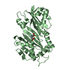
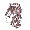
| Title | Endopolygalacturonase from the phytopathogenic fungus Fusarium moniliforme | |||||||||
 Components Components | ENDOPOLYGALACTURONASE Polygalacturonase Polygalacturonase | |||||||||
 Keywords Keywords | HYDROLASE / PECTIN DEGRADATION | |||||||||
| Function / homology |  Function and homology informationendo-polygalacturonase / polygalacturonase activity / cell wall organization / carbohydrate metabolic process / extracellular region Function and homology informationendo-polygalacturonase / polygalacturonase activity / cell wall organization / carbohydrate metabolic process / extracellular regionSimilarity search - Function | |||||||||
| Biological species |  FUSARIUM MONILIFORME (fungus) FUSARIUM MONILIFORME (fungus) | |||||||||
| Method | X-RAY DIFFRACTION / SYNCHROTRON / MIRAS / Resolution: 1.73 Å | |||||||||
 Authors Authors | Federici, L. / Caprari, C. / Mattei, B. / Savino, C. / De Lorenzo, G. / Cervone, F. / Tsernoglou, D. | |||||||||
 Citation Citation | Journal: Proc.Natl.Acad.Sci.USA / Year: 2001 Title: Structural Requirements of Endopolygalacturonase for the Interaction with Pgip (Polygalacturonase-Inhibiting Protein) Authors: Federici, L. / Caprari, C. / Mattei, B. / Savino, C. / Di Matteo, A. / De Lorenzo, G. / Cervone, F. / Tsernoglou, D. #1: Journal: Acta Crystallogr.,Sect.D / Year: 1999 Title: Crystallization and Preliminary X-Ray Diffraction Study of the Endo-Polygalacturonase from Fusarium Moniliforme. Authors: Federici, L. / Mattei, B. / Caprari, C. / Savino, C. / Cervone, F. / Tsernoglou, D. | |||||||||
| History |
|
- Structure visualization
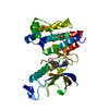
| Structure viewer | Molecule: MolmilJmol/JSmol |
|---|
- Downloads & links
-Download
| PDBx/mmCIF format | 1hg8.cif.gz | 82.7 KB | Display | PDBx/mmCIF format |
|---|---|---|---|---|
| PDB format | pdb1hg8.ent.gz | 62.8 KB | Display | PDB format |
| PDBx/mmJSON format | 1hg8.json.gz | Tree view | PDBx/mmJSON format | |
| Others |  Other downloads Other downloads |
-Validation report
| Arichive directory | https://data.pdbj.org/pub/pdb/validation_reports/hg/1hg8ftp://data.pdbj.org/pub/pdb/validation_reports/hg/1hg8 | HTTPS FTP |
|---|
-Related structure data
| Similar structure data |
|---|


-Links
 PDBj
PDBj- Assembly
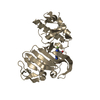
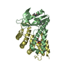
| Deposited unit | 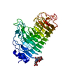
| ||||||||
|---|---|---|---|---|---|---|---|---|---|
| 1 |
| ||||||||
| Unit cell |
|
-Components
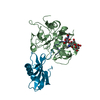
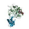
| #1: Protein | Polygalacturonase Mass: 36222.492 Da / Num. of mol.: 1 Source method: isolated from a genetically manipulated source Source: (gene. exp.) FUSARIUM MONILIFORME (fungus) / Production host:  SACCHAROMYCES CEREVISIAE (brewer's yeast) / References: UniProt: Q07181 SACCHAROMYCES CEREVISIAE (brewer's yeast) / References: UniProt: Q07181 |
|---|---|
| #2: Polysaccharide | 2-acetamido-2-deoxy-beta-D-glucopyranose-(1-4)-2-acetamido-2-deoxy-beta-D-glucopyranose / Mass: 424.401 Da / Num. of mol.: 1 Source method: isolated from a genetically manipulated source |
| #3: Sugar | ChemComp-NAG / N-Acetylglucosamine  Type: D-saccharide, beta linking / Mass: 221.208 Da / Num. of mol.: 1 Type: D-saccharide, beta linking / Mass: 221.208 Da / Num. of mol.: 1Source method: isolated from a genetically manipulated source Formula: C8H15NO6 |
| #4: Water | ChemComp-HOH / Water Mass: 18.015 Da / Num. of mol.: 326 / Source method: isolated from a natural source / Formula: H2O Mass: 18.015 Da / Num. of mol.: 326 / Source method: isolated from a natural source / Formula: H2O |
-Experimental details
-Experiment
| Experiment | Method: X-RAY DIFFRACTION / Number of used crystals: 3 |
|---|
- Sample preparation
| Crystal | Density Matthews: 2.6 Å3/Da / Density % sol: 40.9 % | ||||||||||||||||||||||||
|---|---|---|---|---|---|---|---|---|---|---|---|---|---|---|---|---|---|---|---|---|---|---|---|---|---|
| Crystal grow | pH: 4.7 / Details: pH 4.70 | ||||||||||||||||||||||||
| Crystal grow | *PLUS Temperature: 294 K / Method: vapor diffusion, hanging dropDetails: Federici, L., (2001) Acta Crystallogr.,Sect.D, 55, 1359. pH: 4.7 | ||||||||||||||||||||||||
| Components of the solutions | *PLUS
|
-Data collection
| Diffraction | Mean temperature: 100 K |
|---|---|
| Diffraction source | Source: SYNCHROTRON / Site: ELETTRA  / Beamline: 5.2R / Wavelength: 1 / Beamline: 5.2R / Wavelength: 1 |
| Detector | Type: MARRESEARCH / Detector: IMAGE PLATE |
| Radiation | Protocol: SINGLE WAVELENGTH / Monochromatic (M) / Laue (L): M / Scattering type: x-ray |
| Radiation wavelength | Wavelength: 1 Å / Relative weight: 1 |
| Reflection | Resolution: 1.73→17.9 Å / Num. obs: 37710 / % possible obs: 99.4 % / Observed criterion σ(I): 2 / Redundancy: 4.56 % / Rmerge(I) obs: 0.059 |
| Reflection | *PLUS Num. measured all: 204193 |
| Reflection shell | *PLUS % possible obs: 96.6 % / Rmerge(I) obs: 0.302 |
- Processing
| Software |
| |||||||||||||||||||||||||||||||||||||||||||||||||||||||||||||||
|---|---|---|---|---|---|---|---|---|---|---|---|---|---|---|---|---|---|---|---|---|---|---|---|---|---|---|---|---|---|---|---|---|---|---|---|---|---|---|---|---|---|---|---|---|---|---|---|---|---|---|---|---|---|---|---|---|---|---|---|---|---|---|---|---|
| Refinement | Method to determine structure: MIRAS / Resolution: 1.73→17.9 Å / Cross valid method: FREE R-VALUE / σ(F): 2
| |||||||||||||||||||||||||||||||||||||||||||||||||||||||||||||||
| Refinement step | Cycle: LAST / Resolution: 1.73→17.9 Å
| |||||||||||||||||||||||||||||||||||||||||||||||||||||||||||||||
| Refine LS restraints |
| |||||||||||||||||||||||||||||||||||||||||||||||||||||||||||||||
| Software | *PLUS Name: REFMAC / Classification: refinement | |||||||||||||||||||||||||||||||||||||||||||||||||||||||||||||||
| Refinement | *PLUS Rfactor obs: 0.1728 / Rfactor Rfree: 0.203 | |||||||||||||||||||||||||||||||||||||||||||||||||||||||||||||||
| Solvent computation | *PLUS | |||||||||||||||||||||||||||||||||||||||||||||||||||||||||||||||
| Displacement parameters | *PLUS | |||||||||||||||||||||||||||||||||||||||||||||||||||||||||||||||
| Refine LS restraints | *PLUS
|
