 Movie
Movie Controller

[English] 日本語
 Yorodumi
- PDB-1hfc: 1.56 ANGSTROM STRUCTURE OF MATURE TRUNCATED HUMAN FIBROBLAST COLL... -
+ Open data

- Basic information
| Entry | Database: PDB / ID: 1hfc | ||||||
|---|---|---|---|---|---|---|---|
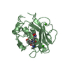
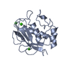
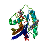
| Title | 1.56 ANGSTROM STRUCTURE OF MATURE TRUNCATED HUMAN FIBROBLAST COLLAGENASE | ||||||
 Components Components | FIBROBLAST COLLAGENASE | ||||||
 Keywords Keywords |  METALLOPROTEASE METALLOPROTEASE | ||||||
| Function / homology |  Function and homology informationinterstitial collagenase / cellular response to UV-A / protein metabolic process / Basigin interactions / Activation of Matrix Metalloproteinases / Collagen degradation / collagen catabolic process / extracellular matrix disassembly / Degradation of the extracellular matrix / extracellular matrix organization ...interstitial collagenase / cellular response to UV-A / protein metabolic process / Basigin interactions / Activation of Matrix Metalloproteinases / Collagen degradation / collagen catabolic process / extracellular matrix disassembly / Degradation of the extracellular matrix / extracellular matrix organization / extracellular matrix / positive regulation of protein-containing complex assembly / metalloendopeptidase activity / Regulation of Insulin-like Growth Factor (IGF) transport and uptake by Insulin-like Growth Factor Binding Proteins (IGFBPs) / peptidase activity / Interleukin-4 and Interleukin-13 signaling / endopeptidase activity / serine-type endopeptidase activity / proteolysis / extracellular space / zinc ion binding / extracellular region Function and homology informationinterstitial collagenase / cellular response to UV-A / protein metabolic process / Basigin interactions / Activation of Matrix Metalloproteinases / Collagen degradation / collagen catabolic process / extracellular matrix disassembly / Degradation of the extracellular matrix / extracellular matrix organization ...interstitial collagenase / cellular response to UV-A / protein metabolic process / Basigin interactions / Activation of Matrix Metalloproteinases / Collagen degradation / collagen catabolic process / extracellular matrix disassembly / Degradation of the extracellular matrix / extracellular matrix organization / extracellular matrix / positive regulation of protein-containing complex assembly / metalloendopeptidase activity / Regulation of Insulin-like Growth Factor (IGF) transport and uptake by Insulin-like Growth Factor Binding Proteins (IGFBPs) / peptidase activity / Interleukin-4 and Interleukin-13 signaling / endopeptidase activity / serine-type endopeptidase activity / proteolysis / extracellular space / zinc ion binding / extracellular regionSimilarity search - Function | ||||||
| Biological species |  Homo sapiens (human) Homo sapiens (human) | ||||||
| Method | X-RAY DIFFRACTION / Resolution: 1.5 Å | ||||||
 Authors Authors | Spurlino, J.C. / Smith, D.L. | ||||||
 Citation Citation | Journal: Proteins / Year: 1994 Title: 1.56 A structure of mature truncated human fibroblast collagenase. Authors: Spurlino, J.C. / Smallwood, A.M. / Carlton, D.D. / Banks, T.M. / Vavra, K.J. / Johnson, J.S. / Cook, E.R. / Falvo, J. / Wahl, R.C. / Pulvino, T.A. / Wendoloski, J.J. / Smith, D.L. #1: Journal: Nat.Struct.Biol. / Year: 1994Title: Structure of Human Neutrophil Collagenase Reveals Large S1' Specificity Pocket Authors: Stams, T. / Spurlino, J.C. / Smith, D.L. / Wahl, R.C. / Ho, T.F. / Qoronfleh, M.W. / Banks, T.M. / Rubin, B. | ||||||
| History |
| ||||||
| Remark 700 | SHEET S1 AS PRESENTED ON SHEET RECORDS BELOW IS A FIVE-STRANDED SHEET. THE HET GROUP HAP IS LOCATED ...SHEET S1 AS PRESENTED ON SHEET RECORDS BELOW IS A FIVE-STRANDED SHEET. THE HET GROUP HAP IS LOCATED IN SUCH A POSITION THAT IT COULD BE CONSIDERED THE SIXTH STRAND OF THIS SHEET SHEET AND PRO 238 - PHE 242 COULD BE CONSIDERED THE SEVENTH STRAND OF THIS SHEET. |
- Structure visualization
| Structure viewer | Molecule: MolmilJmol/JSmol |
|---|
- Downloads & links
-Download
| PDBx/mmCIF format | 1hfc.cif.gz | 48 KB | Display | PDBx/mmCIF format |
|---|---|---|---|---|
| PDB format | pdb1hfc.ent.gz | 32.8 KB | Display | PDB format |
| PDBx/mmJSON format | 1hfc.json.gz | Tree view | PDBx/mmJSON format | |
| Others |  Other downloads Other downloads |
-Validation report
| Arichive directory | https://data.pdbj.org/pub/pdb/validation_reports/hf/1hfcftp://data.pdbj.org/pub/pdb/validation_reports/hf/1hfc | HTTPS FTP |
|---|
-Related structure data
| Similar structure data |
|---|


-Links
 PDBj
PDBj


- Assembly
| Deposited unit | 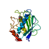
| ||||||||
|---|---|---|---|---|---|---|---|---|---|
| 1 |
| ||||||||
| Unit cell |
| ||||||||
| Atom site foot note | 1: RESIDUES GLU 209 AND TYR 210 FORM A CIS-PEPTIDE BOND. |
-Components
| #1: Protein | Mass: 18865.541 Da / Num. of mol.: 1 Source method: isolated from a genetically manipulated source Source: (gene. exp.) Homo sapiens (human) / References: UniProt: P03956, interstitial collagenase | ||||||
|---|---|---|---|---|---|---|---|
| #2: Chemical |   Mass: 65.409 Da / Num. of mol.: 2 / Source method: obtained synthetically / Formula: Zn Mass: 65.409 Da / Num. of mol.: 2 / Source method: obtained synthetically / Formula: Zn#3: Chemical | ChemComp-CA / |   Mass: 40.078 Da / Num. of mol.: 1 / Source method: obtained synthetically / Formula: Ca Mass: 40.078 Da / Num. of mol.: 1 / Source method: obtained synthetically / Formula: Ca#4: Chemical | ChemComp-PLH / |   Mass: 349.425 Da / Num. of mol.: 1 / Source method: obtained synthetically / Formula: C18H27N3O4 Mass: 349.425 Da / Num. of mol.: 1 / Source method: obtained synthetically / Formula: C18H27N3O4#5: Water | ChemComp-HOH / | Water Mass: 18.015 Da / Num. of mol.: 88 / Source method: isolated from a natural source / Formula: H2O Mass: 18.015 Da / Num. of mol.: 88 / Source method: isolated from a natural source / Formula: H2O |
-Experimental details
-Experiment
| Experiment | Method: X-RAY DIFFRACTION |
|---|
- Sample preparation
| Crystal | Density Matthews: 2.34 Å3/Da / Density % sol: 47.41 % | ||||||||||||||||||||||||||||||||||||||||||
|---|---|---|---|---|---|---|---|---|---|---|---|---|---|---|---|---|---|---|---|---|---|---|---|---|---|---|---|---|---|---|---|---|---|---|---|---|---|---|---|---|---|---|---|
| Crystal grow | *PLUS pH: 7.5 / Method: batch method | ||||||||||||||||||||||||||||||||||||||||||
| Components of the solutions | *PLUS
|
-Data collection
| Radiation | Scattering type: x-ray |
|---|---|
| Radiation wavelength | Relative weight: 1 |
| Reflection | *PLUS Highest resolution: 1.56 Å / Lowest resolution: 2.83 Å / Num. obs: 23912 / % possible obs: 89.7 % / Num. measured all: 64262 / Rmerge(I) obs: 0.0642 |
| Reflection shell | *PLUS Highest resolution: 1.56 Å / Lowest resolution: 1.66 Å / % possible obs: 73.7 % / Num. unique obs: 3218 / Num. measured obs: 5791 / Rmerge(I) obs: 0.2688 |
- Processing
| Software |
| ||||||||||||||||||||||||||||||||||||||||||||||||||||||||||||
|---|---|---|---|---|---|---|---|---|---|---|---|---|---|---|---|---|---|---|---|---|---|---|---|---|---|---|---|---|---|---|---|---|---|---|---|---|---|---|---|---|---|---|---|---|---|---|---|---|---|---|---|---|---|---|---|---|---|---|---|---|---|
| Refinement | Resolution: 1.5→10 Å / σ(F): 2 /
| ||||||||||||||||||||||||||||||||||||||||||||||||||||||||||||
| Refinement step | Cycle: LAST / Resolution: 1.5→10 Å
| ||||||||||||||||||||||||||||||||||||||||||||||||||||||||||||
| Refine LS restraints |
| ||||||||||||||||||||||||||||||||||||||||||||||||||||||||||||
| Software | *PLUS Name: X-PLOR/PROLSQ / Classification: refinement | ||||||||||||||||||||||||||||||||||||||||||||||||||||||||||||
| Refinement | *PLUS Highest resolution: 1.56 Å / Rfactor obs: 0.174 | ||||||||||||||||||||||||||||||||||||||||||||||||||||||||||||
| Solvent computation | *PLUS | ||||||||||||||||||||||||||||||||||||||||||||||||||||||||||||
| Displacement parameters | *PLUS | ||||||||||||||||||||||||||||||||||||||||||||||||||||||||||||
| Refine LS restraints | *PLUS
|
