 Movie
Movie Controller


+ Open data

- Basic information
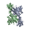
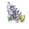
| Entry | Database: PDB / ID: 1h4v | ||||||
|---|---|---|---|---|---|---|---|
| Title | HISTIDYL-TRNA SYNTHETASE from Thermus Thermophilus (ligand free) | ||||||
 Components Components | HISTIDYL-TRNA SYNTHETASE Histidine—tRNA ligase Histidine—tRNA ligase | ||||||
 Keywords Keywords | TRNA SYNTHETASE / CLASS IIA AMINOACYL-TRNA SYNTHETASE / ATP + L-HISTIDINE TRNA(HIS)-> AMP + PPI + L-HISTIDYL-TRNA(HIS) | ||||||
| Function / homology |  Function and homology informationhistidine-tRNA ligase / histidine-tRNA ligase activity / histidyl-tRNA aminoacylation / ATP binding / cytoplasm Function and homology informationhistidine-tRNA ligase / histidine-tRNA ligase activity / histidyl-tRNA aminoacylation / ATP binding / cytoplasmSimilarity search - Function | ||||||
| Biological species |   THERMUS THERMOPHILUS (bacteria) THERMUS THERMOPHILUS (bacteria) | ||||||
| Method | X-RAY DIFFRACTION / SYNCHROTRON / MOLECULAR REPLACEMENT / Resolution: 2.4 Å | ||||||
 Authors Authors | Cusack, S. / Yaremchuk, A. / Tukalo, M. | ||||||
 Citation Citation | Journal: J.Mol.Biol. / Year: 2001 Title: A Succession of Substrate Induced Conformational Changes Ensures the Amino Acid Specificity of Thermus Thermophilus Prolyl-tRNA Synthetase: Comparison with Histidyl-tRNA Synthetase Authors: Yaremchuk, A. / Tukalo, M. / Grotli, M. / Cusack, S. #1: Journal: Biochemistry / Year: 1997Title: Crystal Structure Analysis of the Activation of Histidine by Thermus Thermophilus Histidyl-tRNA Synthetase Authors: Aberg, A. / Yaremchuk, A. / Tukalo, M. / Rasmussen, B. / Cusack, S. | ||||||
| History |
|
- Structure visualization
| Structure viewer | Molecule: MolmilJmol/JSmol |
|---|
- Downloads & links
-Download
| PDBx/mmCIF format | 1h4v.cif.gz | 92.9 KB | Display | PDBx/mmCIF format |
|---|---|---|---|---|
| PDB format | pdb1h4v.ent.gz | 70.2 KB | Display | PDB format |
| PDBx/mmJSON format | 1h4v.json.gz | Tree view | PDBx/mmJSON format | |
| Others |  Other downloads Other downloads |
-Validation report
| Arichive directory | https://data.pdbj.org/pub/pdb/validation_reports/h4/1h4vftp://data.pdbj.org/pub/pdb/validation_reports/h4/1h4v | HTTPS FTP |
|---|
-Related structure data
| Related structure data | 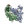 1h4qC  1h4sC 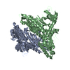 1h4tC  1hc7C  1adjS S: Starting model for refinement C: citing same article ( |
|---|---|
| Similar structure data |


-Links
 PDBj
PDBj
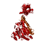
- Assembly
| Deposited unit | 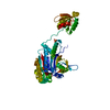
| ||||||||
|---|---|---|---|---|---|---|---|---|---|
| 1 | 
| ||||||||
| Unit cell |
|
-Components
| #1: Protein | Histidine—tRNA ligase Mass: 47125.918 Da / Num. of mol.: 1 / Source method: isolated from a natural source / Details: LIGAND FREE FORM OF THE ENZYME / Source: (natural) THERMUS THERMOPHILUS (bacteria) / Strain: HB27References: UniProt: P56194, UniProt: P62374*PLUS, histidine-tRNA ligase |
|---|---|
| #2: Chemical | ChemComp-SO4 / Sulfate  Mass: 96.063 Da / Num. of mol.: 1 / Source method: obtained synthetically / Formula: SO4 Mass: 96.063 Da / Num. of mol.: 1 / Source method: obtained synthetically / Formula: SO4 |
| #3: Water | ChemComp-HOH / Water Mass: 18.015 Da / Num. of mol.: 65 / Source method: isolated from a natural source / Formula: H2O Mass: 18.015 Da / Num. of mol.: 65 / Source method: isolated from a natural source / Formula: H2O |
| Sequence details | THE SEQUENCE OF T. THERMOPHILUS HISTIDYL-TRNA SYNTHETASE IS GIVEN IN BIOCHEMISTRY 36, 3084 - 3094, ...THE SEQUENCE OF T. THERMOPHIL |
-Experimental details
-Experiment
| Experiment | Method: X-RAY DIFFRACTION / Number of used crystals: 1 |
|---|
- Sample preparation
| Crystal | Density Matthews: 2.76 Å3/Da / Density % sol: 55.4 % | ||||||||||||||||||||||||||||||||||||||||||||||||||||||
|---|---|---|---|---|---|---|---|---|---|---|---|---|---|---|---|---|---|---|---|---|---|---|---|---|---|---|---|---|---|---|---|---|---|---|---|---|---|---|---|---|---|---|---|---|---|---|---|---|---|---|---|---|---|---|---|
| Crystal grow | pH: 7 / Details: DESCRIBED IN REFERENCE 1., pH 7.00 | ||||||||||||||||||||||||||||||||||||||||||||||||||||||
| Crystal grow | *PLUS Temperature: 295 K / pH: 7.6 / Method: vapor diffusion, hanging drop | ||||||||||||||||||||||||||||||||||||||||||||||||||||||
| Components of the solutions | *PLUS
|
-Data collection
| Diffraction | Mean temperature: 100 K |
|---|---|
| Diffraction source | Source: SYNCHROTRON / Site: ESRF  / Beamline: ID2 / Wavelength: 0.99 / Beamline: ID2 / Wavelength: 0.99 |
| Detector | Type: MARRESEARCH / Detector: IMAGE PLATE / Date: Apr 15, 1997 / Details: MIRRORS |
| Radiation | Monochromator: SI CRYSTAL / Protocol: SINGLE WAVELENGTH / Monochromatic (M) / Laue (L): M / Scattering type: x-ray |
| Radiation wavelength | Wavelength: 0.99 Å / Relative weight: 1 |
| Reflection | Resolution: 2.4→12 Å / Num. obs: 39592 / % possible obs: 86.7 % / Observed criterion σ(I): 0 / Redundancy: 2.2 % / Rmerge(I) obs: 0.074 |
| Reflection shell | Resolution: 2.4→2.46 Å / Redundancy: 1.8 % / Rmerge(I) obs: 0.14 / Mean I/σ(I) obs: 4.9 / % possible all: 72.1 |
| Reflection | *PLUS Num. obs: 17744 / Num. measured all: 39592 |
| Reflection shell | *PLUS % possible obs: 72.1 % / Rmerge(I) obs: 0.137 |
- Processing
| Software |
| ||||||||||||||||||||||||||||||||||||||||||||||||||||||||||||||||||||||||||||||||
|---|---|---|---|---|---|---|---|---|---|---|---|---|---|---|---|---|---|---|---|---|---|---|---|---|---|---|---|---|---|---|---|---|---|---|---|---|---|---|---|---|---|---|---|---|---|---|---|---|---|---|---|---|---|---|---|---|---|---|---|---|---|---|---|---|---|---|---|---|---|---|---|---|---|---|---|---|---|---|---|---|---|
| Refinement | Method to determine structure: MOLECULAR REPLACEMENT Starting model: 1ADJ Resolution: 2.4→12 Å / Isotropic thermal model: RESTRAINED / Cross valid method: THROUGHOUT / σ(F): 0 Details: RESIDUES 57-62 AND 117-120 ARE DISORDERED. OTHER RESIDUES WITH SIDE-CHAIN ATOMS WITH ZERO OCCUPANCY ARE POORLY ORDERED.
| ||||||||||||||||||||||||||||||||||||||||||||||||||||||||||||||||||||||||||||||||
| Solvent computation | Solvent model: FLAT MODEL / Bsol: 45.1 Å2 / ksol: 0.44 e/Å3 | ||||||||||||||||||||||||||||||||||||||||||||||||||||||||||||||||||||||||||||||||
| Displacement parameters | Biso mean: 32.7 Å2
| ||||||||||||||||||||||||||||||||||||||||||||||||||||||||||||||||||||||||||||||||
| Refinement step | Cycle: LAST / Resolution: 2.4→12 Å
| ||||||||||||||||||||||||||||||||||||||||||||||||||||||||||||||||||||||||||||||||
| Refine LS restraints |
| ||||||||||||||||||||||||||||||||||||||||||||||||||||||||||||||||||||||||||||||||
| Xplor file |
|
