 Movie
Movie Controller

[English] 日本語
 Yorodumi
- PDB-1h1c: Histidinol-phosphate aminotransferase (HisC) from Thermotoga maritima -
+ Open data

- Basic information
| Entry | Database: PDB / ID: 1h1c | ||||||
|---|---|---|---|---|---|---|---|
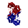
| Title | Histidinol-phosphate aminotransferase (HisC) from Thermotoga maritima | ||||||
 Components Components | HISTIDINOL-PHOSPHATE AMINOTRANSFERASE | ||||||
 Keywords Keywords |  TRANSFERASE / AMINOTRANSFERASE / HISTIDINE BIOSYNTHESIS TRANSFERASE / AMINOTRANSFERASE / HISTIDINE BIOSYNTHESIS | ||||||
| Function / homology |  Function and homology informationhistidinol-phosphate transaminase / histidinol-phosphate transaminase activity / L-histidine biosynthetic process / pyridoxal phosphate binding / protein homodimerization activity Function and homology informationhistidinol-phosphate transaminase / histidinol-phosphate transaminase activity / L-histidine biosynthetic process / pyridoxal phosphate binding / protein homodimerization activitySimilarity search - Function | ||||||
| Biological species |   THERMOTOGA MARITIMA (bacteria) THERMOTOGA MARITIMA (bacteria) | ||||||
| Method | X-RAY DIFFRACTION / SYNCHROTRON / MAD / Resolution: 2.85 Å | ||||||
 Authors Authors | Vega, M.C. / Fernandez, F.J. / Wilmanns, M. | ||||||
 Citation Citation | Journal: J.Biol.Chem. / Year: 2004 Title: Structural Studies of the Catalytic Reaction Pathway of a Hyperthermophilic Histidinol-Phosphate Aminotransferase Authors: Fernandez, F.J. / Vega, M.C. / Lehmann, F. / Sandmeier, E. / Gehring, H. / Christen, P. / Wilmanns, M. #1: Journal: J.Mol.Biol. / Year: 2001Title: Crystal Structure of Histidinol Phosphate Aminotransferase (Hisc) from Escherichia Coli, and its Covalent Complex with Pyridoxal-5'-Phosphate and L-Histidinol Phosphate Authors: Sivaraman, J. / Li, Y. / Larocque, R. / Schrag, J.D. / Cygler, M. / Matte, A. | ||||||
| History |
|
- Structure visualization
| Structure viewer | Molecule: MolmilJmol/JSmol |
|---|
- Downloads & links
-Download
| PDBx/mmCIF format | 1h1c.cif.gz | 268.3 KB | Display | PDBx/mmCIF format |
|---|---|---|---|---|
| PDB format | pdb1h1c.ent.gz | 227.8 KB | Display | PDB format |
| PDBx/mmJSON format | 1h1c.json.gz | Tree view | PDBx/mmJSON format | |
| Others |  Other downloads Other downloads |
-Validation report
| Arichive directory | https://data.pdbj.org/pub/pdb/validation_reports/h1/1h1cftp://data.pdbj.org/pub/pdb/validation_reports/h1/1h1c | HTTPS FTP |
|---|
-Related structure data


-Links
 PDBj
PDBj- Assembly
| Deposited unit | 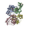
| ||||||||||||||||
|---|---|---|---|---|---|---|---|---|---|---|---|---|---|---|---|---|---|
| 1 | 
| ||||||||||||||||
| 2 | 
| ||||||||||||||||
| Unit cell |
| ||||||||||||||||
| Noncrystallographic symmetry (NCS) | NCS oper:
|
-Components
| #1: Protein | Mass: 39772.996 Da / Num. of mol.: 4 Source method: isolated from a genetically manipulated source Details: RESIDUE 202 LYSINE-PYRIDOXAL-5'-PHOSPHATE / Source: (gene. exp.) THERMOTOGA MARITIMA (bacteria) / Plasmid: PETM11 / Production host: ESCHERICHIA COLI (E. coli) / Strain (production host): B834(DE3)References: UniProt: Q9X0D0, histidinol-phosphate transaminase#2: Chemical | ChemComp-PLP / Pyridoxal phosphate  Mass: 247.142 Da / Num. of mol.: 4 / Source method: obtained synthetically / Formula: C8H10NO6P Mass: 247.142 Da / Num. of mol.: 4 / Source method: obtained synthetically / Formula: C8H10NO6P#3: Water | ChemComp-HOH / | Water Mass: 18.015 Da / Num. of mol.: 133 / Source method: isolated from a natural source / Formula: H2O Mass: 18.015 Da / Num. of mol.: 133 / Source method: isolated from a natural source / Formula: H2OCompound details | CONVERTS L-HISTIDINOL-PHOSPHATE + 2-OXOGLUTARATE TO 3- (IMIDAZOL-4-YL)-2-OXOPROPYL PHOSPHATE + L- ...CONVERTS L-HISTIDINOL | |
|---|
-Experimental details
-Experiment
| Experiment | Method: X-RAY DIFFRACTION / Number of used crystals: 1 |
|---|
- Sample preparation
| Crystal | Density Matthews: 2.53 Å3/Da / Density % sol: 51 % | ||||||||||||||||||||||||||||||||||||||||||
|---|---|---|---|---|---|---|---|---|---|---|---|---|---|---|---|---|---|---|---|---|---|---|---|---|---|---|---|---|---|---|---|---|---|---|---|---|---|---|---|---|---|---|---|
| Crystal grow | pH: 7 / Details: pH 7.00 | ||||||||||||||||||||||||||||||||||||||||||
| Crystal grow | *PLUS Temperature: 20 ℃ / pH: 7.5 / Method: vapor diffusion, sitting drop | ||||||||||||||||||||||||||||||||||||||||||
| Components of the solutions | *PLUS
|
-Data collection
| Diffraction | Mean temperature: 100 K | ||||||||||||
|---|---|---|---|---|---|---|---|---|---|---|---|---|---|
| Diffraction source | Source: SYNCHROTRON / Site: EMBL/DESY, HAMBURG  / Beamline: BW7A / Wavelength: 0.9778,0.9862,0.9184 / Beamline: BW7A / Wavelength: 0.9778,0.9862,0.9184 | ||||||||||||
| Detector | Type: MARRESEARCH / Detector: CCD / Date: Dec 15, 2001 | ||||||||||||
| Radiation | Protocol: MAD / Monochromatic (M) / Laue (L): M / Scattering type: x-ray | ||||||||||||
| Radiation wavelength |
| ||||||||||||
| Reflection | Resolution: 2.85→20 Å / Num. obs: 29624 / % possible obs: 99.8 % / Redundancy: 4.1 % / Rmerge(I) obs: 0.092 / Net I/σ(I): 6.98 | ||||||||||||
| Reflection shell | Resolution: 2.85→2.95 Å / Redundancy: 3.3 % / Rmerge(I) obs: 0.381 / Mean I/σ(I) obs: 2.38 / % possible all: 95.7 | ||||||||||||
| Reflection | *PLUS Highest resolution: 2.85 Å / Lowest resolution: 20 Å |
- Processing
| Software |
| ||||||||||||||||||||||||||||||||||||||||||||||||||||||||||||
|---|---|---|---|---|---|---|---|---|---|---|---|---|---|---|---|---|---|---|---|---|---|---|---|---|---|---|---|---|---|---|---|---|---|---|---|---|---|---|---|---|---|---|---|---|---|---|---|---|---|---|---|---|---|---|---|---|---|---|---|---|---|
| Refinement | Method to determine structure: MAD / Resolution: 2.85→20 Å / Cross valid method: THROUGHOUT / σ(F): 0
| ||||||||||||||||||||||||||||||||||||||||||||||||||||||||||||
| Displacement parameters | Biso mean: 21.92 Å2
| ||||||||||||||||||||||||||||||||||||||||||||||||||||||||||||
| Refinement step | Cycle: LAST / Resolution: 2.85→20 Å
| ||||||||||||||||||||||||||||||||||||||||||||||||||||||||||||
| Refine LS restraints |
| ||||||||||||||||||||||||||||||||||||||||||||||||||||||||||||
| Refine LS restraints NCS | NCS model details: RESTRAINTS | ||||||||||||||||||||||||||||||||||||||||||||||||||||||||||||
| LS refinement shell | Resolution: 2.85→2.914 Å / Total num. of bins used: 20 /
| ||||||||||||||||||||||||||||||||||||||||||||||||||||||||||||
| Refinement | *PLUS Lowest resolution: 20 Å / % reflection Rfree: 9.3 % / Rfactor Rwork: 0.22 | ||||||||||||||||||||||||||||||||||||||||||||||||||||||||||||
| Solvent computation | *PLUS | ||||||||||||||||||||||||||||||||||||||||||||||||||||||||||||
| Displacement parameters | *PLUS |
