 Movie
Movie Controller

[English] 日本語
 Yorodumi
- PDB-1gyj: The Crystal Structure of YdcE, a 4-Oxalocrotonate Tautomerase Hom... -
+ Open data

- Basic information
| Entry | Database: PDB / ID: 1gyj | ||||||
|---|---|---|---|---|---|---|---|
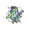
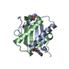
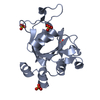
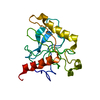
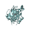
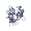
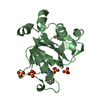
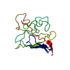
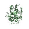
| Title | The Crystal Structure of YdcE, a 4-Oxalocrotonate Tautomerase Homologue from Escherichia coli, Confirms the Structural Basis for Oligomer Diversity | ||||||
 Components Components | HYPOTHETICAL PROTEIN YDCE | ||||||
 Keywords Keywords | ISOMERASE / TAUTOMERASE / HYPOTHETICAL PROTEIN | ||||||
| Function / homology |  Function and homology information Function and homology informationintramolecular oxidoreductase activity, interconverting keto- and enol-groups / Isomerases; Intramolecular oxidoreductases; Interconverting keto- and enol-groups / : / protein homodimerization activity / cytoplasm Similarity search - Function | ||||||
| Biological species |  | ||||||
| Method |  X-RAY DIFFRACTION / MOLECULAR REPLACEMENT / Resolution: 2.1 Å X-RAY DIFFRACTION / MOLECULAR REPLACEMENT / Resolution: 2.1 Å | ||||||
 Authors Authors | Almrud, J. / Kern, A. / Wang, S. / Czerwinski, R. / Johnson, W. / Murzin, A. / Hackert, M. / Whitman, C. | ||||||
 Citation Citation | Journal: Biochemistry / Year: 2002 Title: The Crystal Structure of Ydce, a 4-Oxalocrotonate Tautomerase Homologue from Escherichia Coli, Confirms the Structural Basis for Oligomer Diversity Authors: Almrud, J. / Kern, A. / Wang, S. / Czerwinski, R. / Johnson, W. / Murzin, A. / Hackert, M. / Whitman, C. | ||||||
| History |
|
- Structure visualization
| Structure viewer | Molecule: MolmilJmol/JSmol |
|---|
- Downloads & links
-Download
| PDBx/mmCIF format | 1gyj.cif.gz | 44.1 KB | Display | PDBx/mmCIF format |
|---|---|---|---|---|
| PDB format | pdb1gyj.ent.gz | 31.4 KB | Display | PDB format |
| PDBx/mmJSON format | 1gyj.json.gz | Tree view | PDBx/mmJSON format | |
| Others |  Other downloads Other downloads |
-Validation report
| Summary document | 1gyj_validation.pdf.gz | 433.3 KB | Display | wwPDB validaton report |
|---|---|---|---|---|
| Full document | 1gyj_full_validation.pdf.gz | 435.1 KB | Display | |
| Data in XML | 1gyj_validation.xml.gz | 9.7 KB | Display | |
| Data in CIF | 1gyj_validation.cif.gz | 13.1 KB | Display | |
| Arichive directory | https://data.pdbj.org/pub/pdb/validation_reports/gy/1gyjftp://data.pdbj.org/pub/pdb/validation_reports/gy/1gyj | HTTPS FTP |
-Related structure data


-Links
 PDBj
PDBj- Assembly
| Deposited unit | 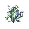
| ||||||||
|---|---|---|---|---|---|---|---|---|---|
| 1 |
| ||||||||
| Unit cell |
| ||||||||
| Noncrystallographic symmetry (NCS) | NCS oper: (Code: given Matrix: (0.161249, -0.963495, 0.213719), Vector: |
-Components
| #1: Protein | Mass: 8550.729 Da / Num. of mol.: 2 Source method: isolated from a genetically manipulated source Source: (gene. exp.) #2: Water | ChemComp-HOH / |  Mass: 18.015 Da / Num. of mol.: 151 / Source method: isolated from a natural source / Formula: H2O Mass: 18.015 Da / Num. of mol.: 151 / Source method: isolated from a natural source / Formula: H2O |
|---|
-Experimental details
-Experiment
| Experiment | Method: X-RAY DIFFRACTION / Number of used crystals: 1 |
|---|
- Sample preparation
| Crystal | Density Matthews: 2.5 Å3/Da / Density % sol: 51 % | ||||||||||||||||||
|---|---|---|---|---|---|---|---|---|---|---|---|---|---|---|---|---|---|---|---|
| Crystal grow | pH: 7.3 Details: PROTEIN BUFFERED IN 50MM SODIUM PHOSPHATE, PH 7.3 PRECIPITANT: 40% SODIUM CITRATE PH 8.5 ITTING DROP: MIX 5UL PROT. WITH 5UL 40% SODIUM CITRATE | ||||||||||||||||||
| Crystal grow | *PLUS Temperature: 4 ℃ / pH: 8.5 / Method: vapor diffusion, hanging drop | ||||||||||||||||||
| Components of the solutions | *PLUS
|
-Data collection
| Diffraction | Mean temperature: 110 K |
|---|---|
| Diffraction source | Source: ROTATING ANODE / Type: RIGAKU RU200 / Wavelength: 1.5418 |
| Detector | Type: MARRESEARCH / Detector: IMAGE PLATE / Date: Dec 15, 2001 / Details: MIRRORS |
| Radiation | Protocol: SINGLE WAVELENGTH / Monochromatic (M) / Laue (L): M / Scattering type: x-ray |
| Radiation wavelength | Wavelength: 1.5418 Å / Relative weight: 1 |
| Reflection | Resolution: 2.1→30 Å / Num. obs: 10344 / % possible obs: 99.7 % / Observed criterion σ(I): 3 / Redundancy: 16 % / Biso Wilson estimate: 27.1 Å2 / Rmerge(I) obs: 0.092 / Net I/σ(I): 11.5 |
| Reflection shell | Resolution: 2.1→2.18 Å / Redundancy: 10 % / Rmerge(I) obs: 0.408 / Mean I/σ(I) obs: 3 / % possible all: 99.1 |
| Reflection | *PLUS Lowest resolution: 30 Å / Num. measured all: 169053 |
| Reflection shell | *PLUS Highest resolution: 2.1 Å / % possible obs: 99.1 % / Rmerge(I) obs: 0.435 |
- Processing
| Software |
| ||||||||||||||||||||||||||||||||||||||||||||||||||||||||||||
|---|---|---|---|---|---|---|---|---|---|---|---|---|---|---|---|---|---|---|---|---|---|---|---|---|---|---|---|---|---|---|---|---|---|---|---|---|---|---|---|---|---|---|---|---|---|---|---|---|---|---|---|---|---|---|---|---|---|---|---|---|---|
| Refinement | Method to determine structure: MOLECULAR REPLACEMENT Starting model: UNPUBLISHED 1.35 ANG. RES. YDCE STRUCTURE DETERMINED BY MIR Resolution: 2.1→30 Å / Cross valid method: THROUGHOUT / σ(F): 2
| ||||||||||||||||||||||||||||||||||||||||||||||||||||||||||||
| Solvent computation | Solvent model: COMBINATION | ||||||||||||||||||||||||||||||||||||||||||||||||||||||||||||
| Displacement parameters | Biso mean: 25.5 Å2
| ||||||||||||||||||||||||||||||||||||||||||||||||||||||||||||
| Refinement step | Cycle: LAST / Resolution: 2.1→30 Å
| ||||||||||||||||||||||||||||||||||||||||||||||||||||||||||||
| Refine LS restraints |
| ||||||||||||||||||||||||||||||||||||||||||||||||||||||||||||
| Refine LS restraints NCS | NCS model details: UNRESTRAINED | ||||||||||||||||||||||||||||||||||||||||||||||||||||||||||||
| LS refinement shell | Resolution: 2.1→2.18 Å / Total num. of bins used: 9
| ||||||||||||||||||||||||||||||||||||||||||||||||||||||||||||
| Refine LS restraints | *PLUS
| ||||||||||||||||||||||||||||||||||||||||||||||||||||||||||||
| LS refinement shell | *PLUS Highest resolution: 2.1 Å |
