 Movie
Movie Controller

[English] 日本語
 Yorodumi
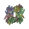
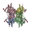
- PDB-1gr0: myo-inositol 1-phosphate synthase from Mycobacterium tuberculosis... -
+ Open data

- Basic information
| Entry | Database: PDB / ID: 1gr0 | ||||||
|---|---|---|---|---|---|---|---|
| Title | myo-inositol 1-phosphate synthase from Mycobacterium tuberculosis in complex with NAD and zinc. | ||||||
 Components Components | INOSITOL-3-PHOSPHATE SYNTHASE | ||||||
 Keywords Keywords | ISOMERASE / OXIDOREDUCTASE / PSI / PROTEIN STRUCTURE INITIATIVE / TB STRUCTURAL GENOMICS CONSORTIUM / TB / TBSGC | ||||||
| Function / homology |  Function and homology information Function and homology informationMycothiol biosynthesis / inositol-3-phosphate synthase / mycothiol biosynthetic process / inositol-3-phosphate synthase activity / inositol biosynthetic process / phospholipid biosynthetic process / : / zinc ion binding / cytosolSimilarity search - Function | ||||||
| Biological species |   MYCOBACTERIUM TUBERCULOSIS (bacteria) MYCOBACTERIUM TUBERCULOSIS (bacteria) | ||||||
| Method | X-RAY DIFFRACTION / SYNCHROTRON / MOLECULAR REPLACEMENT / Resolution: 1.95 Å | ||||||
 Authors Authors | Norman, R.A. / Murray-Rust, J. / McDonald, N.Q. / TB Structural Genomics Consortium (TBSGC) | ||||||
 Citation Citation | Journal: Structure / Year: 2002 Title: Crystal Structure of Inositol 1-Phosphate Synthase from Mycobacterium Tuberculosis, a Key Enzyme in Phosphatidylinositol Synthesis Authors: Norman, R.A. / Mcalister, M.S.B. / Murray-Rust, J. / Movahedzadeh, F. / Stoker, N.G. / Mcdonald, N.Q. | ||||||
| History |
| ||||||
| Remark 700 | SHEET THE SHEET STRUCTURE OF THIS MOLECULE IS BIFURCATED. IN ORDER TO REPRESENT THIS FEATURE IN ... SHEET THE SHEET STRUCTURE OF THIS MOLECULE IS BIFURCATED. IN ORDER TO REPRESENT THIS FEATURE IN THE SHEET RECORDS BELOW, TWO SHEETS ARE DEFINED. |
- Structure visualization
| Structure viewer | Molecule: MolmilJmol/JSmol |
|---|
- Downloads & links
-Download
| PDBx/mmCIF format | 1gr0.cif.gz | 81.7 KB | Display | PDBx/mmCIF format |
|---|---|---|---|---|
| PDB format | pdb1gr0.ent.gz | 59.9 KB | Display | PDB format |
| PDBx/mmJSON format | 1gr0.json.gz | Tree view | PDBx/mmJSON format | |
| Others |  Other downloads Other downloads |
-Validation report
| Arichive directory | https://data.pdbj.org/pub/pdb/validation_reports/gr/1gr0ftp://data.pdbj.org/pub/pdb/validation_reports/gr/1gr0 | HTTPS FTP |
|---|
-Related structure data
| Similar structure data | |
|---|---|
| Other databases |


-Links
 PDBj
PDBj


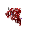
- Assembly
| Deposited unit | 
| ||||||||
|---|---|---|---|---|---|---|---|---|---|
| 1 | 
| ||||||||
| Unit cell |
|
-Components
| #1: Protein | / IPS / MYO-INOSITOL-1-PHOSPHATE SYNTHASE / MI-1-P SYNTHASE / MIP SYNTHASE / HYPOTHETICAL 40.1 KDA ...IPS / MYO-INOSITOL-1-PHOSPHATE SYNTHASE / MI-1-P SYNTHASE / MIP SYNTHASE / HYPOTHETICAL 40.1 KDA PROTEIN RV0046C Mass: 40139.492 Da / Num. of mol.: 1 Source method: isolated from a genetically manipulated source Source: (gene. exp.) MYCOBACTERIUM TUBERCULOSIS (bacteria) / Plasmid: PET-15B / Production host: ESCHERICHIA COLI (E. coli)References: UniProt: P71703, UniProt: P9WKI1*PLUS, inositol-3-phosphate synthase |
|---|---|
| #2: Chemical | ChemComp-NAD / Nicotinamide adenine dinucleotide  Mass: 663.425 Da / Num. of mol.: 1 / Source method: obtained synthetically / Formula: C21H27N7O14P2 / Comment: NAD*YM Mass: 663.425 Da / Num. of mol.: 1 / Source method: obtained synthetically / Formula: C21H27N7O14P2 / Comment: NAD*YM |
| #3: Chemical | ChemComp-ZN /   Mass: 65.409 Da / Num. of mol.: 1 / Source method: obtained synthetically / Formula: Zn Mass: 65.409 Da / Num. of mol.: 1 / Source method: obtained synthetically / Formula: Zn |
| #4: Chemical | ChemComp-CAC / Cacodylic acid  Mass: 136.989 Da / Num. of mol.: 1 / Source method: obtained synthetically / Formula: C2H6AsO2 Mass: 136.989 Da / Num. of mol.: 1 / Source method: obtained synthetically / Formula: C2H6AsO2 |
| #5: Water | ChemComp-HOH / Water Mass: 18.015 Da / Num. of mol.: 170 / Source method: isolated from a natural source / Formula: H2O Mass: 18.015 Da / Num. of mol.: 170 / Source method: isolated from a natural source / Formula: H2O |
| Compound details | THE ENZYME INOSITOL-1-PHOSPHATE SYNTHASE IS PART OF THE PHOSPHATIDYLINOSITOL BIOSYNTHESIS PATHWAY ...THE ENZYME INOSITOL-1-PHOSPHATE SYNTHASE IS PART OF THE PHOSPHATID |
-Experimental details
-Experiment
| Experiment | Method: X-RAY DIFFRACTION / Number of used crystals: 1 |
|---|
- Sample preparation
| Crystal | Density Matthews: 2.8 Å3/Da / Density % sol: 56 % | ||||||||||||||||||||||||||||||||||||||||||||||||
|---|---|---|---|---|---|---|---|---|---|---|---|---|---|---|---|---|---|---|---|---|---|---|---|---|---|---|---|---|---|---|---|---|---|---|---|---|---|---|---|---|---|---|---|---|---|---|---|---|---|
| Crystal grow | pH: 7 Details: OPTIMAL CRYSTALLISATION CONDITIONS: PEG 4000 (6.2-6.7% W/V), SODIUM CACODYLATE (50MM, PH7.0), CALCIUM ACETATE (100MM)., pH 7.00 | ||||||||||||||||||||||||||||||||||||||||||||||||
| Crystal grow | *PLUS Method: vapor diffusion, hanging drop | ||||||||||||||||||||||||||||||||||||||||||||||||
| Components of the solutions | *PLUS
|
-Data collection
| Diffraction | Mean temperature: 100 K |
|---|---|
| Diffraction source | Source: SYNCHROTRON / Site: ESRF  / Beamline: ID14-4 / Wavelength: 0.979 / Beamline: ID14-4 / Wavelength: 0.979 |
| Detector | Type: ADSC CCD / Detector: CCD / Date: Sep 15, 2000 / Details: MIRRORS |
| Radiation | Monochromator: DOUBLE CRYSTAL / Protocol: SINGLE WAVELENGTH / Monochromatic (M) / Laue (L): M / Scattering type: x-ray |
| Radiation wavelength | Wavelength: 0.979 Å / Relative weight: 1 |
| Reflection | Resolution: 1.95→24.79 Å / Num. obs: 29486 / % possible obs: 90 % / Redundancy: 2.7 % / Biso Wilson estimate: 16.1 Å2 / Rmerge(I) obs: 0.071 / Net I/σ(I): 7.7 |
| Reflection shell | Resolution: 1.95→1.98 Å / Redundancy: 2.6 % / Rmerge(I) obs: 0.338 / Mean I/σ(I) obs: 2.8 / % possible all: 87 |
| Reflection | *PLUS % possible obs: 89.9 % / Num. measured all: 80189 |
| Reflection shell | *PLUS % possible obs: 86.6 % |
- Processing
| Software |
| ||||||||||||||||||||||||||||||||||||||||||||||||||||||||||||||||||||||||||||||||
|---|---|---|---|---|---|---|---|---|---|---|---|---|---|---|---|---|---|---|---|---|---|---|---|---|---|---|---|---|---|---|---|---|---|---|---|---|---|---|---|---|---|---|---|---|---|---|---|---|---|---|---|---|---|---|---|---|---|---|---|---|---|---|---|---|---|---|---|---|---|---|---|---|---|---|---|---|---|---|---|---|---|
| Refinement | Method to determine structure: MOLECULAR REPLACEMENT Starting model: PARTIALLY REFINED MODEL OBTAINED FROM MAD PHASES Resolution: 1.95→24.79 Å / Rfactor Rfree error: 0.006 / Data cutoff high absF: 3021712.68 / Data cutoff low absF: 0 / Isotropic thermal model: RESTRAINED / Cross valid method: THROUGHOUT / σ(F): 0 Details: THE SIDE CHAINS FOR THE FOLLOWING RESIDUES WERE DISORDERED IN ELECTRON DENSITY: THR A 14, ARG A 60, ASP A 268
| ||||||||||||||||||||||||||||||||||||||||||||||||||||||||||||||||||||||||||||||||
| Solvent computation | Solvent model: FLAT MODEL / Bsol: 46.409 Å2 / ksol: 0.382561 e/Å3 | ||||||||||||||||||||||||||||||||||||||||||||||||||||||||||||||||||||||||||||||||
| Displacement parameters | Biso mean: 30.3 Å2
| ||||||||||||||||||||||||||||||||||||||||||||||||||||||||||||||||||||||||||||||||
| Refine analyze |
| ||||||||||||||||||||||||||||||||||||||||||||||||||||||||||||||||||||||||||||||||
| Refinement step | Cycle: LAST / Resolution: 1.95→24.79 Å
| ||||||||||||||||||||||||||||||||||||||||||||||||||||||||||||||||||||||||||||||||
| Refine LS restraints |
| ||||||||||||||||||||||||||||||||||||||||||||||||||||||||||||||||||||||||||||||||
| LS refinement shell | Resolution: 1.95→2.07 Å / Rfactor Rfree error: 0.022 / Total num. of bins used: 6
| ||||||||||||||||||||||||||||||||||||||||||||||||||||||||||||||||||||||||||||||||
| Xplor file |
| ||||||||||||||||||||||||||||||||||||||||||||||||||||||||||||||||||||||||||||||||
| Refinement | *PLUS % reflection Rfree: 5 % | ||||||||||||||||||||||||||||||||||||||||||||||||||||||||||||||||||||||||||||||||
| Solvent computation | *PLUS | ||||||||||||||||||||||||||||||||||||||||||||||||||||||||||||||||||||||||||||||||
| Displacement parameters | *PLUS | ||||||||||||||||||||||||||||||||||||||||||||||||||||||||||||||||||||||||||||||||
| Refine LS restraints | *PLUS
|
