 Movie
Movie Controller

[English] 日本語
 Yorodumi
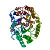
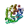
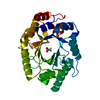
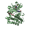
- PDB-1gok: Thermostable xylanase I from Thermoascus aurantiacus- Crystal form II -
+ Open data

- Basic information
| Entry | Database: PDB / ID: 1gok | ||||||||||||
|---|---|---|---|---|---|---|---|---|---|---|---|---|---|
| Title | Thermostable xylanase I from Thermoascus aurantiacus- Crystal form II | ||||||||||||
 Components Components | ENDO-1,4-BETA-XYLANASE Xylanase Xylanase | ||||||||||||
 Keywords Keywords | HYDROLASE / XYLANASE / FAMILY 10 / PLANT CELL WALL DEGRADATION | ||||||||||||
| Function / homology |  Function and homology informationendo-1,4-beta-xylanase activity / endo-1,4-beta-xylanase / xylan catabolic process Function and homology informationendo-1,4-beta-xylanase activity / endo-1,4-beta-xylanase / xylan catabolic processSimilarity search - Function | ||||||||||||
| Biological species |  THERMOASCUS AURANTIACUS (fungus) THERMOASCUS AURANTIACUS (fungus) | ||||||||||||
| Method | X-RAY DIFFRACTION / SYNCHROTRON / MOLECULAR REPLACEMENT / Resolution: 1.14 Å | ||||||||||||
 Authors Authors | Lo Leggio, L. / Pickersgill, R.W. | ||||||||||||
 Citation Citation | Journal: FEBS Lett. / Year: 2001 Title: Substrate Specificity and Subsite Mobility in T. Aurantiacus Xylanase 10A Authors: Lo Leggio, L. / Kalogiannis, S. / Eckert, K. / Teixeira, S.C.M. / Bhat, M.K. / Andrei, C. / Pickersgill, R.W. / Larsen, S. #1: Journal: Acta Crystallogr.,Sect.D / Year: 2001 Title: Anisotropic Refinement of the Structure of Thermoascus Aurantiacus Xylanase I Authors: Teixeira, S. / Lo Leggio, L. / Pickersgill, R. / Cardin, C. #2: Journal: Proteins: Struct.,Funct., Genet. / Year: 1999 Title: High Resolution Structure and Sequence of T. Aurantiacus Xylanase I: Implications for the Evolution of Thermostability in Family 10 Xylanases and Enzymes with Beta/Alpha Barrel Architecture Authors: Lo Leggio, L. / Kalogiannis, S. / Bhat, M.K. / Pickersgill, R.W. #3: Journal: Biochem.Soc.Trans. / Year: 1998 Title: Superfamilies: The 4/7 Superfamily of Beta/ Alpha-Barrel Glycosidases and the Right-Handed Parallel Beta-Helix Superfamily Authors: Pickersgill, R. / Harris, G. / Lo Leggio, L. / Mayans, O. / Jenkins, J. #4: Journal: Structural Studies of Xylanases and Endoglucanases / Year: 1997Title: Structure Solution of Thermoascus Aurantiacus Xylanase. Structural Studies of Xylanases and Endoglucanases Authors: Lo Leggio, L. | ||||||||||||
| History |
| ||||||||||||
| Remark 700 | SHEET DETERMINATION METHOD: DSSP THE SHEETS PRESENTED AS "AA" IN EACH CHAIN ON SHEET RECORDS BELOW ... SHEET DETERMINATION METHOD: DSSP THE SHEETS PRESENTED AS "AA" IN EACH CHAIN ON SHEET RECORDS BELOW IS ACTUALLY AN 10-STRANDED BARREL THIS IS REPRESENTED BY A 11-STRANDED SHEET IN WHICH THE FIRST AND LAST STRANDS ARE IDENTICAL. |
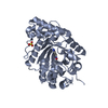
- Structure visualization
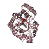
| Structure viewer | Molecule: MolmilJmol/JSmol |
|---|
- Downloads & links
-Download
| PDBx/mmCIF format | 1gok.cif.gz | 78 KB | Display | PDBx/mmCIF format |
|---|---|---|---|---|
| PDB format | pdb1gok.ent.gz | 56.3 KB | Display | PDB format |
| PDBx/mmJSON format | 1gok.json.gz | Tree view | PDBx/mmJSON format | |
| Others |  Other downloads Other downloads |
-Validation report
| Arichive directory | https://data.pdbj.org/pub/pdb/validation_reports/go/1gokftp://data.pdbj.org/pub/pdb/validation_reports/go/1gok | HTTPS FTP |
|---|
-Related structure data
| Related structure data | 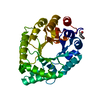 1gomC 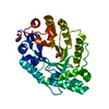 1gooC  1goqC 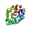 1gorC  1k6aC 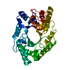 2exoS C: citing same article ( S: Starting model for refinement |
|---|---|
| Similar structure data |


-Links
 PDBj
PDBj
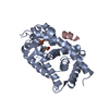
- Assembly
| Deposited unit | 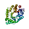
| ||||||||
|---|---|---|---|---|---|---|---|---|---|
| 1 |
| ||||||||
| Unit cell |
|
-Components
| #1: Protein | Xylanase / XYLANASE / 1 / 4-BETA-D-XYLAN XYLANOHYDROLASE / TAXI Mass: 32890.711 Da / Num. of mol.: 1 / Source method: isolated from a natural source / Source: (natural) THERMOASCUS AURANTIACUS (fungus) / References: UniProt: P23360, endo-1,4-beta-xylanase |
|---|---|
| #2: Water | ChemComp-HOH / Water Mass: 18.015 Da / Num. of mol.: 248 / Source method: isolated from a natural source / Formula: H2O Mass: 18.015 Da / Num. of mol.: 248 / Source method: isolated from a natural source / Formula: H2O |
| Sequence details | RESIDUES 1-26 REFER TO THE SIGNAL PEPTIDE. IT IS NOT KNOWN IF GLN 303 IS PRESENT IN THE CRYSTAL |
-Experimental details
-Experiment
| Experiment | Method: X-RAY DIFFRACTION / Number of used crystals: 2 |
|---|
- Sample preparation
| Crystal | Density Matthews: 2.02 Å3/Da / Density % sol: 32.9 % / Description: THE PH OF CRYSTALLIZATION WAS NOT BUFFERED |
|---|---|
| Crystal grow | Method: vapor diffusion, hanging drop / pH: 7 Details: HANGING DROPS CONTAINING 1:1 RATIO OF 20 MG/ML PROTEIN SOLUTION AND RESERVOIR SOLUTION (12 % TO 25 % PEG 6,000)., pH 7.00 |
-Data collection
| Diffraction | Mean temperature: 293 K |
|---|---|
| Diffraction source | Source: SYNCHROTRON / Site: SRS  / Beamline: PX9.6 / Wavelength: 0.87 / Beamline: PX9.6 / Wavelength: 0.87 |
| Detector | Type: MARRESEARCH / Detector: IMAGE PLATE / Date: Aug 20, 1995 / Details: MIRRORS |
| Radiation | Monochromator: SILICON / Protocol: SINGLE WAVELENGTH / Monochromatic (M) / Laue (L): M / Scattering type: x-ray |
| Radiation wavelength | Wavelength: 0.87 Å / Relative weight: 1 |
| Reflection | Resolution: 1.14→39.5 Å / Num. obs: 80691 / % possible obs: 85.2 % / Observed criterion σ(I): 0 / Redundancy: 2.9 % / Biso Wilson estimate: 10.8 Å2 / Rmerge(I) obs: 0.078 / Net I/σ(I): 9.82 |
| Reflection shell | Resolution: 1.14→1.15 Å / Redundancy: 2.1 % / Rmerge(I) obs: 0.238 / Mean I/σ(I) obs: 3.15 / % possible all: 78.8 |
- Processing
| Software |
| ||||||||||||||||||||||||||||||||||||||||||||||||||||||||||||||||||||||||||||||||
|---|---|---|---|---|---|---|---|---|---|---|---|---|---|---|---|---|---|---|---|---|---|---|---|---|---|---|---|---|---|---|---|---|---|---|---|---|---|---|---|---|---|---|---|---|---|---|---|---|---|---|---|---|---|---|---|---|---|---|---|---|---|---|---|---|---|---|---|---|---|---|---|---|---|---|---|---|---|---|---|---|---|
| Refinement | Method to determine structure: MOLECULAR REPLACEMENT Starting model: PDB ENTRY 2EXO Resolution: 1.14→39.5 Å / Rfactor Rfree error: 0.004 / Data cutoff high absF: 100000 / Isotropic thermal model: RESTRAINED / Cross valid method: THROUGHOUT / σ(F): 0 / Stereochemistry target values: MLF Details: A VERY IMPORTANT STEP TOWARDS STRUCTURE SOLUTION WAS REFINEMENT USING ARP (AUTOMATED REFINEMENT PROCEDURE)
| ||||||||||||||||||||||||||||||||||||||||||||||||||||||||||||||||||||||||||||||||
| Solvent computation | Solvent model: FLAT MODEL / Bsol: 56.6 Å2 / ksol: 0.372 e/Å3 | ||||||||||||||||||||||||||||||||||||||||||||||||||||||||||||||||||||||||||||||||
| Displacement parameters | Biso mean: 13.87 Å2
| ||||||||||||||||||||||||||||||||||||||||||||||||||||||||||||||||||||||||||||||||
| Refine analyze |
| ||||||||||||||||||||||||||||||||||||||||||||||||||||||||||||||||||||||||||||||||
| Refinement step | Cycle: LAST / Resolution: 1.14→39.5 Å
| ||||||||||||||||||||||||||||||||||||||||||||||||||||||||||||||||||||||||||||||||
| Refine LS restraints |
| ||||||||||||||||||||||||||||||||||||||||||||||||||||||||||||||||||||||||||||||||
| LS refinement shell | Resolution: 1.14→1.16 Å / Rfactor Rfree error: 0.038 / Total num. of bins used: 20
| ||||||||||||||||||||||||||||||||||||||||||||||||||||||||||||||||||||||||||||||||
| Xplor file | Serial no: 1 / Param file: PROTEIN_REP.PARAM / Topol file: PROTEIN.TOP |
